Ann Rehabil Med.
2016 Feb;40(1):28-33. 10.5535/arm.2016.40.1.28.
Clinical Usefulness of Sonoelastography in Infants With Congenital Muscular Torticollis
- Affiliations
-
- 1Department of Rehabilitation Medicine, Daegu Fatima Hospital, Daegu, Korea. zilee@hanmail.net
- 2Department of Radiology, Daegu Fatima Hospital, Daegu, Korea.
- KMID: 2155162
- DOI: http://doi.org/10.5535/arm.2016.40.1.28
Abstract
OBJECTIVE
To evaluate the clinical usefulness of sonoelastography in infants with congenital muscular torticollis (CMT).
METHODS
The medical records of 215 infants clinically diagnosed with CMT were retrospectively reviewed. Fifty-three infants met the inclusion criteria as follows: 1) infants diagnosed as CMT with a palpable neck mass before 3 months of age, 2) infants who were evaluated initially by both B-mode ultrasonography and sonoelastography, and 3) infants who had received physical therapy after being diagnosed with CMT. We checked the thickness of the sternocleidomastoid (SCM) muscles in B-mode ultrasonography, strain ratio of the SCM muscles in sonoelastography, and treatment duration. We evaluated the correlation between the treatment duration and the following factors: SCM muscle thickness, ratio of SCM muscle thickness on the affected to unaffected side (A/U ratio), and strain ratio.
RESULTS
Both the thickness of the affected SCM muscle and the A/U ratio did not show significant correlation with the treatment duration (p=0.66, p=0.90). The strain ratio of the affected SCM muscle was significantly greater than that of the unaffected SCM muscle (p<0.001), and the strain ratio showed significant correlation with the treatment duration (p=0.001).
CONCLUSION
Sonoelastography may be a useful adjunctive tool to B-mode ultrasonography for evaluating infants with CMT, especially when predicting their rehabilitation outcomes.
MeSH Terms
Figure
-
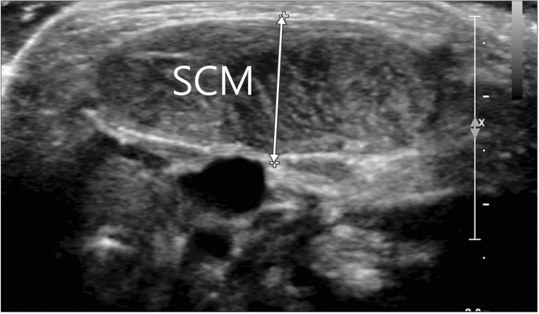
Fig. 1 Transverse view of the sternocleidomastoid (SCM) muscle in B-mode ultrasonogram. White arrow indicates thickness of the SCM muscle.
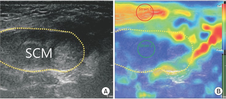
Fig. 2 Transverse view of sternocleidomastoid (SCM) muscle in B-mode ultrasonogram (A) and sonoelastogram (B). Each yellow dotted circle is the cross-sectional area of SCM muscle. In sonoelastogram (B), the colors ranging from red (soft) to blue (hard) represent relative stiffness of the tissue. Red circle (strain 1) indicates region of interest (ROI) of subcutaneous tissue (reference tissue), and green circle (strain 2) is ROI of SCM muscle.
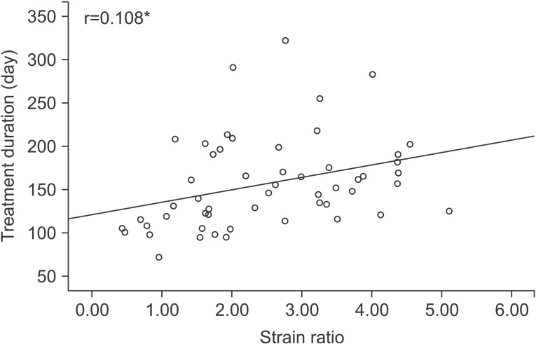
Fig. 3 Correlation between strain ratio and treatment duration in a scatter plot. *p<0.01, Spearman correlation test.
Reference
-
1. Ling CM, Low YS. Sternomastoid tumor and muscular torticollis. Clin Orthop Relat Res. 1972; 86:144–150. PMID: 5065412.
Article2. Cheng JC, Au AW. Infantile torticollis: a review of 624 cases. J Pediatr Orthop. 1994; 14:802–808. PMID: 7814599.3. Porter SB, Blount BW. Pseudotumor of infancy and congenital muscular torticollis. Am Fam Physician. 1995; 52:1731–1736. PMID: 7484683.4. Yim SY, Lee IY, Park MC, Kim JH. Differential diagnosis and management of abnormal posture of the head and neck. J Korean Med Assoc. 2009; 52:705–718.
Article5. Cheng JC, Tang SP, Chen TM. Sternocleidomastoid pseudotumor and congenital muscular torticollis in infants: a prospective study of 510 cases. J Pediatr. 1999; 134:712–716. PMID: 10356139.
Article6. Brans J, Aramideh M, Bosch A, Speelman H. Late presentation of congenital muscular torticollis: a nondystonic cause of torticollis. J Neurol. 1996; 243:354–356. PMID: 8965110.
Article7. Singer C, Green BA, Bruce JH, Bowen BC, Weiner WJ. Late presentation of congenital muscular torticollis: use of MR imaging and CT scan in diagnosis. Mov Disord. 1994; 9:100–103. PMID: 8139587.
Article8. Yu CC, Wong FH, Lo LJ, Chen YR. Craniofacial deformity in patients with uncorrected congenital muscular torticollis: an assessment from three-dimensional computed tomography imaging. Plast Reconstr Surg. 2004; 113:24–33. PMID: 14707619.
Article9. Lee JY, Koh SE, Lee IS, Jung H, Lee J, Kang JI, et al. The cervical range of motion as a factor affecting outcome in patients with congenital muscular torticollis. Ann Rehabil Med. 2013; 37:183–190. PMID: 23705112.
Article10. Jung AY, Kang EY, Lee SH, Nam DH, Cheon JH, Kim HJ. Factors that affect the rehabilitation duration in patients with congenital muscular torticollis. Ann Rehabil Med. 2015; 39:18–24. PMID: 25750867.
Article11. Han SJ, Shin BM, Lee JM, Yoon TS. Factors affecting rehabilitation outcome of congenital muscular torticollis. J Korean Acad Rehabil Med. 2010; 34:643–649.12. Han JD, Kim SH, Lee SJ, Park MC, Yim SY. The thickness of the sternocleidomastoid muscle as a prognostic factor for congenital muscular torticollis. Ann Rehabil Med. 2011; 35:361–368. PMID: 22506145.
Article13. Ophir J, Cespedes I, Ponnekanti H, Yazdi Y, Li X. Elastography: a quantitative method for imaging the elasticity of biological tissues. Ultrason Imaging. 1991; 13:111–134. PMID: 1858217.
Article14. Niitsu M, Michizaki A, Endo A, Takei H, Yanagisawa O. Muscle hardness measurement by using ultrasound elastography: a feasibility study. Acta Radiol. 2011; 52:99–105. PMID: 21498334.
Article15. Ariji Y, Katsumata A, Hiraiwa Y, Izumi M, Iida Y, Goto M, et al. Use of sonographic elastography of the masseter muscles for optimizing massage pressure: a preliminary study. J Oral Rehabil. 2009; 36:627–635. PMID: 19602100.
Article16. Kim MO, Kim SJ. Results of the conservative management in congenital musculartorticollis. J Korean Acad Rehabil Med. 1992; 16:42–50.17. Do TT. Congenital muscular torticollis: current concepts and review of treatment. Curr Opin Pediatr. 2006; 18:26–29. PMID: 16470158.18. Lee YT, Yoon K, Kim YB, Chung PW, Hwang JH, Park YS, et al. Clinical features and outcome of physiotherapy in early presenting congenital muscular torticollis with severe fibrosis on ultrasonography: a prospective study. J Pediatr Surg. 2011; 46:1526–1531. PMID: 21843719.
Article19. Drakonaki EE, Allen GM. Magnetic resonance imaging, ultrasound and real-time ultrasound elastography of the thigh muscles in congenital muscle dystrophy. Skeletal Radiol. 2010; 39:391–396. PMID: 20205351.
Article20. Sconfienza LM, Silvestri E, Orlandi D, Fabbro E, Ferrero G, Martini C, et al. Real-time sonoelastography of the plantar fascia: comparison between patients with plantar fasciitis and healthy control subjects. Radiology. 2013; 267:195–200. PMID: 23297327.
Article21. Klauser AS, Miyamoto H, Tamegger M, Faschingbauer R, Moriggl B, Klima G, et al. Achilles tendon assessed with sonoelastography: histologic agreement. Radiology. 2013; 267:837–842. PMID: 23449953.
Article
- Full Text Links
-
- Actions
-
Cited
- CITED
-
- Close
- Share
-
- Similar articles
-
- Familial Congenital Muscular Torticollis: A Case Report
- Two Cases of Sternomastoid Tumor
- Unipolar Release of the Sternocleidomastoideus in Congenital Muscular Torticollis in Children
- Congenital Torticollis with Bilateral Sternocleidomastoid Muscle Contracture
- Ultrasonographic Measurement of the Sternocleidomastoid Muscle in Congenital Muscular Torticollis
