Facial and occlusal esthetic improvements of an adult skeletal Class III malocclusion using surgical, orthodontic, and implant treatment
- Affiliations
-
- 1Department of Orthodontics, University of Sagrado Coracao, Bauru, Sao Paulo, Brazil.
- 2Department of Diagnosis and Surgery, School of Dentistry at Araraquara, Univ Estadual Paulista - UNESP, Araraquara, Sao Paulo, Brazil. molon.foar@yahoo.com.br
- 3Department of Dental Materials and Prosthodontics, School of Dentistry at Araraquara, Univ Estadual Paulista - UNESP, Araraquara, Sao Paulo, Brazil.
- 4Department of Oral Implantology, University of Sagrado Coracao - USC, Bauru, Sao Paulo, Brazil.
- KMID: 2152897
- DOI: http://doi.org/10.4041/kjod.2016.46.1.42
Abstract
- The aim of this clinical report is to describe the complex treatment of an adult Class III malocclusion patient who was disappointed with the outcome of a previous oral rehabilitation. Interdisciplinary treatment planning was performed with a primary indication for implant removal because of marginal bone loss and gingival recession, followed by orthodontic and surgical procedures to correct the esthetics and skeletal malocclusion. The comprehensive treatment approach included: (1) implant removal in the area of the central incisors; (2) combined orthodontic decompensation with mesial displacement and forced extrusion of the lateral incisors; (3) extraction of the lateral incisors and placement of new implants corresponding to the central incisors, which received provisional crowns; (4) orthognathic surgery for maxillary advancement to improve occlusal and facial relationships; and finally, (5) orthodontic refinement followed by definitive prosthetic rehabilitation of the maxillary central incisors and reshaping of the adjacent teeth. At the three-year follow-up, clinical and radiographic examinations showed successful replacement of the central incisors and improved skeletal and esthetic appearances. Moreover, a Class II molar relationship was obtained with an ideal overbite, overjet, and intercuspation. In conclusion, we report the successful esthetic anterior rehabilitation of a complex case in which interdisciplinary treatment planning improved facial harmony, provided gingival architecture with sufficient width and thickness, and improved smile esthetics, resulting in enhanced patient comfort and satisfaction. This clinical case report might be useful to improve facial esthetics and occlusion in patients with dentoalveolar and skeletal defects.
Keyword
MeSH Terms
Figure
-
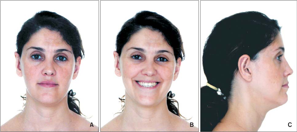
Figure 1 Facial photographs obtained before treatment.

Figure 2 Pretreatment photographs. A-E, Intraoral photographs obtained before treatment; F-J, pretreatment dental casts.
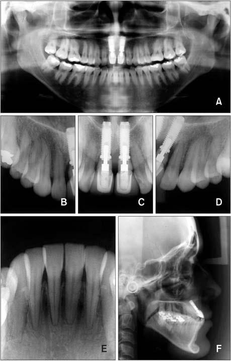
Figure 3 Pretreatment panoramic, periapical, and lateral cephalometric radiographs.
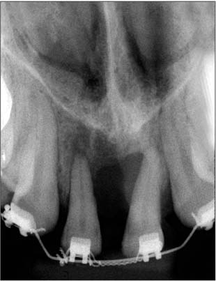
Figure 4 A periapical radiograph obtained after mesial displacement and extrusion of the lateral incisors.
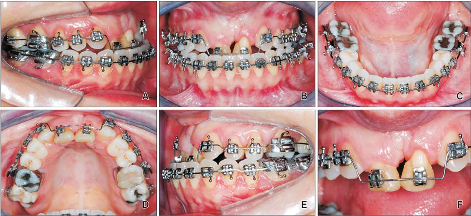
Figure 5 Intraoral photographs taken after extrusion of the lateral incisors.
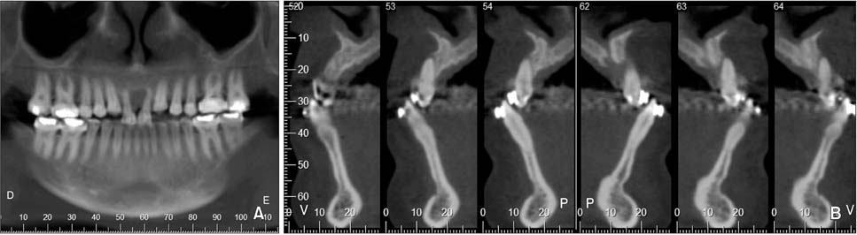
Figure 6 A cone-beam computed tomography scan acquired prior to implant placement. A, Panoramic coronal view; B, sequential parasagittal views.
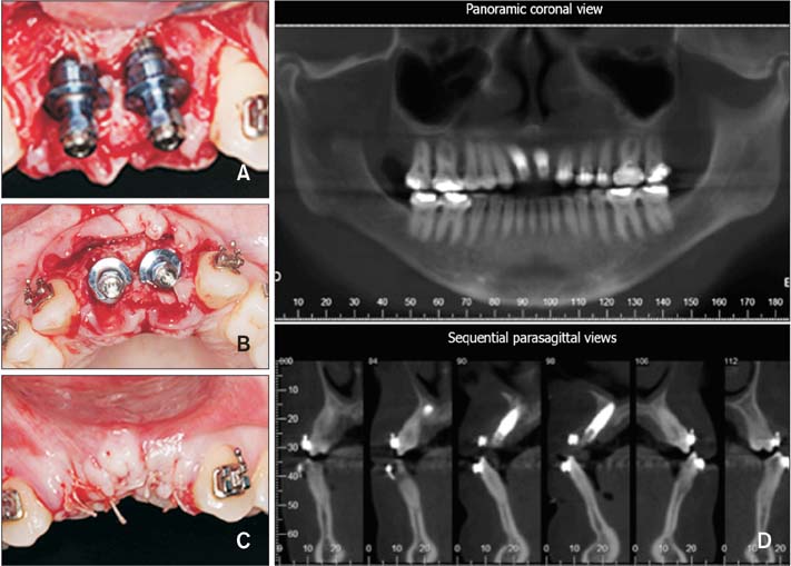
Figure 7 Implant installation. A-C, Intraoral photographs of implant placement; D, a cone-beam computed tomography scan acquired after implant installation.
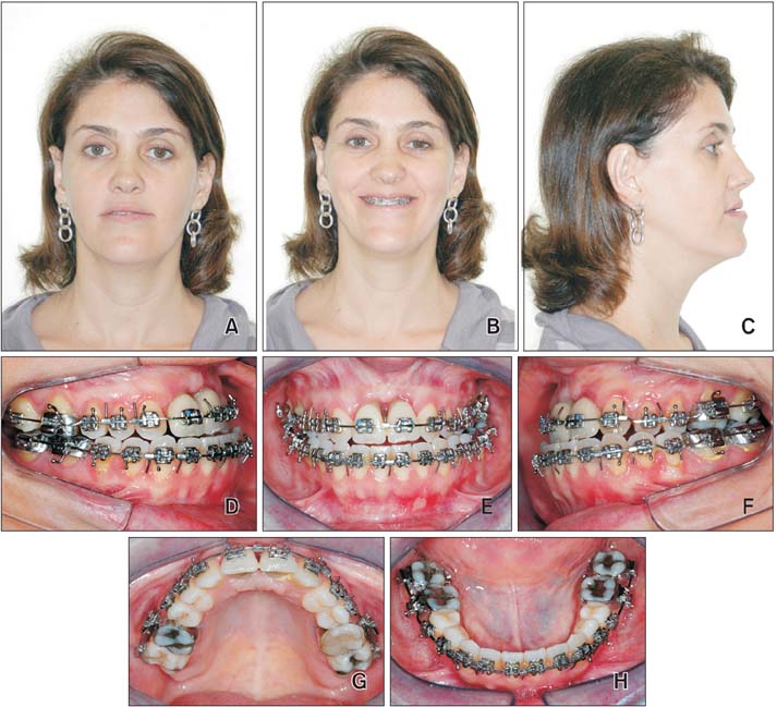
Figure 8 The photographs taken after orthodontic decompensation. A-C, Facial photographs; D-H, intraoral photographs.
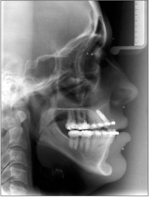
Figure 9 A lateral cephalometric radiograph obtained after orthodontic decompensation.
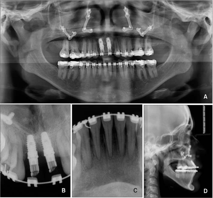
Figure 10 Final panoramic, periapical, and lateral cephalometric radiographs obtained after orthognathic surgery.
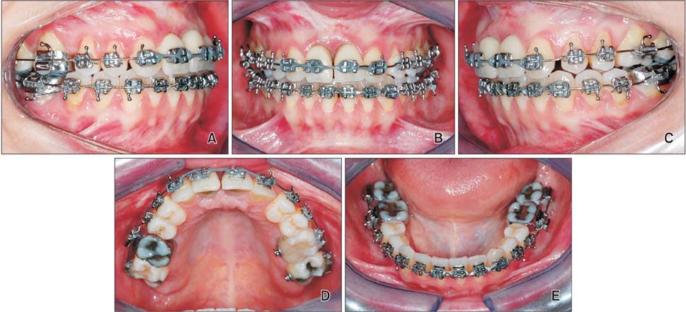
Figure 11 Intraoral photographs taken after orthodontic movement and finalization of the surgical procedures.

Figure 12 Final photographs after orthodontic appliance removal. A-C, Facial photographs; D-H, intraoral photographs.
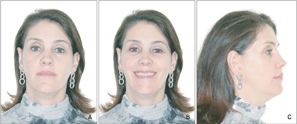
Figure 13 Facial photographs taken at the three-year follow-up.
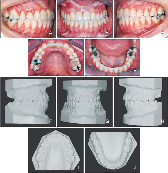
Figure 14 The follow up after three years. A-E, Intraoral photographs taken three years post-treatment; F-J, final treatment dental casts.

Figure 15 The cephalometric superimposition. A, Pre-treatment (black line) and post-decompensation (blue line). B, Pretreatment (black line) and post-treatment (red line).
Reference
-
1. de Molon RS, Avila ED, Cirelli JA, Mollo-Jr FD, Andrade MF, Barros-Filho LA, et al. A combined approach for the treatment of resorbed fresh sockets allowing immediate implant restoration. A 2-year follow-up. J Oral Implantol. 2015; 41:712–718.
Article2. Choquet V, Hermans M, Adriaenssens P, Daelemans P, Tarnow DP, Malevez C. Clinical and radiographic evaluation of the papilla level adjacent to single-tooth dental implants. A retrospective study in the maxillary anterior region. J Periodontol. 2001; 72:1364–1371.
Article3. Kan JY, Rungcharassaeng K. Interimplant papilla preservation in the esthetic zone: a report of six consecutive cases. Int J Periodontics Restorative Dent. 2003; 23:249–259.4. Belser UC, Schmid B, Higginbottom F, Buser D. Outcome analysis of implant restorations located in the anterior maxilla: a review of the recent literature. Int J Oral Maxillofac Implants. 2004; 19:Suppl. 30–42.5. Spear FM, Kokich VG, Mathews DP. Interdisciplinary management of anterior dental esthetics. J Am Dent Assoc. 2006; 137:160–169.
Article6. de Molon RS, Morais-Camilo JA, Verzola MH, Faeda RS, Pepato MT, Marcantonio E Jr. Impact of diabetes mellitus and metabolic control on bone healing around osseointegrated implants: removal torque and histomorphometric analysis in rats. Clin Oral Implants Res. 2013; 24:831–837.
Article7. de Molon RS, de Avila ED, Cirelli JA, Cardoso Mde A, Capelozza-Filho L, Borelli Barros LA. Optimizing maxillary aesthetics of a severe compromised tooth through orthodontic movement and dental implants. Case Rep Dent. 2014; 2014:103808.
Article8. de Avila ÉD, de Molon RS, de Assis Mollo F Jr, de Barros LA, Capelozza Filho L, de Almeida Cardoso M, et al. Multidisciplinary approach for the aesthetic treatment of maxillary lateral incisors agenesis: thinking about implants? Oral Surg Oral Med Oral Pathol Oral Radiol. 2012; 114:e22–e28.
Article9. de Molon RS, de Avila ED, de Barros-Filho LA, Ricci WA, Tetradis S, Cirelli JA, et al. Reconstruction of the alveolar buccal bone plate in compromised fresh socket after immediate implant placement followed by immediate provisionalization. J Esthet Restor Dent. 2015; 27:122–135.
Article10. Buser D, Martin W, Belser UC. Optimizing esthetics for implant restorations in the anterior maxilla: anatomic and surgical considerations. Int J Oral Maxillofac Implants. 2004; 19:Suppl. 43–61.11. Fürhauser R, Florescu D, Benesch T, Haas R, Mailath G, Watzek G. Evaluation of soft tissue around single-tooth implant crowns: the pink esthetic score. Clin Oral Implants Res. 2005; 16:639–644.
Article12. Jung RE, Pjetursson BE, Glauser R, Zembic A, Zwahlen M, Lang NP. A systematic review of the 5-year survival and complication rates of implant-supported single crowns. Clin Oral Implants Res. 2008; 19:119–130.
Article13. Kan JY, Rungcharassaeng K, Fillman M, Caruso J. Tissue architecture modification for anterior implant esthetics: an interdisciplinary approach. Eur J Esthet Dent. 2009; 4:104–117.14. Nisapakultorn K, Suphanantachat S, Silkosessak O, Rattanamongkolgul S. Factors affecting soft tissue level around anterior maxillary single-tooth implants. Clin Oral Implants Res. 2010; 21:662–670.
Article15. Papalexiou V, Novaes AB Jr, Ribeiro RF, Muglia V, Oliveira RR. Influence of the interimplant distance on crestal bone resorption and bone density: a histomorphometric study in dogs. J Periodontol. 2006; 77:614–621.
Article16. de Barros LA, de Almeida Cardoso M, de Avila ED, de Molon RS, Siqueira DF, Mollo-Junior Fde A, et al. Six-year follow-up of maxillary anterior rehabilitation with forced orthodontic extrusion: achieving esthetic excellence with a multidisciplinary approach. Am J Orthod Dentofacial Orthop. 2013; 144:607–615.
Article17. de Molon RS, de Avila ÉD, de Souza JA, Nogueira AV, Cirelli CC, Cirelli JA. Combination of orthodontic movement and periodontal therapy for full root coverage in a Miller class III recession: a case report with 12 years of follow-up. Braz Dent J. 2012; 23:758–763.
Article18. de Molon RS, de Avila ED, de Souza JA, Nogueira AV, Cirelli CC, Margonar R, et al. Forced orthodontic eruption for augmentation of soft and hard tissue prior to implant placement. Contemp Clin Dent. 2013; 4:243–247.
Article19. de Molon RS, Kim YJ, Dos Santos-Pinto A, Cirelli JA. Improvement of an anterior infrabone defect using combined periodontal and orthodontic therapy: A 6-year follow-up case report. Eur J Dent. 2014; 8:407–411.
Article20. Corrente G, Abundo R, Re S, Cardaropoli D, Cardaropoli G. Orthodontic movement into infrabony defects in patients with advanced periodontal disease: a clinical and radiological study. J Periodontol. 2003; 74:1104–1109.
Article21. Korayem M, Flores-Mir C, Nassar U, Olfert K. Implant site development by orthodontic extrusion. A systematic review. Angle Orthod. 2008; 78:752–760.22. Sonego CL, Bobrowski ÂN, Chagas OL Jr, Torriani MA. Aesthetic and functional implications following rotation of the maxillomandibular complex in orthognathic surgery: a systematic review. Int J Oral Maxillofac Surg. 2014; 43:40–45.
Article23. Tarnow DP, Magner AW, Fletcher P. The effect of the distance from the contact point to the crest of bone on the presence or absence of the interproximal dental papilla. J Periodontol. 1992; 63:995–996.
Article24. Funato A, Salama MA, Ishikawa T, Garber DA, Salama H. Timing, positioning, and sequential staging in esthetic implant therapy: a four-dimensional perspective. Int J Periodontics Restorative Dent. 2007; 27:313–323.25. Garber DA, Belser UC. Restoration-driven implant placement with restoration-generated site development. Compend Contin Educ Dent. 1995; 16:796798–802. 80426. Palacci P. Aesthetic treatment of the anterior maxilla: soft and hard tissue considerations. Oral Maxillofac Surg Clin North Am. 2004; 16:127–137.
Article27. Seibert JS, Salama H. Alveolar ridge preservation and reconstruction. Periodontol 2000. 1996; 11:69–84.
Article28. Margonar R, Queiroz TP, Marcantonio É, Luvizuto ER, Faloni AP, Betoni W Jr, et al. Rehabilitation of the maxillary arch after bone graft using immediate loading with implant-supported fixed restoration. J Craniofac Surg. 2014; 25:e44–e48.
Article29. Milinkovic I, Cordaro L. Are there specific indications for the different alveolar bone augmentation procedures for implant placement? A systematic review. Int J Oral Maxillofac Surg. 2014; 43:606–625.
Article30. Margonar R, Queiroz TP, Luvizuto ER, Santos PL, Wady AF, Paleari AG. Anterior tooth rehabilitation with frozen homogenous bone graft and immediately loaded titanium implant using computer-guided surgery. J Craniofac Surg. 2012; 23:e470–e472.
Article31. Shiu YT, Weiss JA, Hoying JB, Iwamoto MN, Joung IS, Quam CT. The role of mechanical stresses in angiogenesis. Crit Rev Biomed Eng. 2005; 33:431–510.
Article32. de Ávila ED, de Molon RS, Loffredo LC, Massucato EM, Hochuli-Vieira E. Health-related quality of life and depression in patients with dentofacial deformity. Oral Maxillofac Surg. 2013; 17:187–191.
Article
- Full Text Links
-
- Actions
-
Cited
- CITED
-
- Close
- Share
-
- Similar articles
-
- Orthodontic Treatment Using Invisalign First® in Pediatric Patients with Mild Skeletal Malocclusion: Case Reports
- Class III nonsurgical treatment using indirect skeletal anchorage: A case report
- A study on the vertical dysplasia in the skeletal class iii malocclusion
- A study of the characteristics of craniofacial skeleton on orthognathic surgical cases with skeletal Class III malocclusion
- Morphology of mandibular symphysis and positioning of lower incisors in the skeletal Class III malocclusions
