Usefulness of Digital Tomosynthesis for the Detection of Airway Obstruction: A Case Report of Bronchial Carcinosarcoma
- Affiliations
-
- 1Department of Radiology, Korea University Ansan Hospital, Korea University of College of Medicine, Ansan, Korea. quellquell@naver.com
- 2Department of Pulmonology, Korea University Ansan Hospital, Korea University of College of Medicine, Ansan, Korea.
- 3Department of Pathology, Korea University Ansan Hospital, Korea University of College of Medicine, Ansan, Korea.
- KMID: 2148507
- DOI: http://doi.org/10.4143/crt.2013.220
Abstract
- Bronchial carcinosarcoma is a very rare malignant tumor that is composed of carcinomatous and sarcomatous elements. We describe the first case in which digital tomosynthesis was useful for the evaluation of airway obstruction by bronchial carcinosarcoma that was overlooked on initial chest radiography.
Keyword
Figure
-
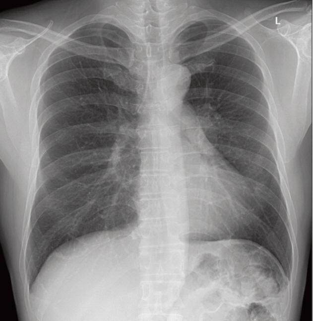
Fig. 1. Initial chest radiography. The endobronchial mass with left upper lobar bronchial narrowing was overlooked on the initial chest radiography.

Fig. 2. Digital tomosynthesis (DTS). (A) DTS reveals a lobulating contoured endobronchial lesion nearly obstructing the left main bronchus. (B) The tumor extends to the left upper lobar bronchus and the left lower lobar bronchus. However, the superior segmental bronchus is not obliterated on the last image with patent three holes (arrows). The extent and margins of the endobronchial tumor are well demarcated on serial anterior-to-posterior coronal images from the DTS. The yellow overlay and gray patent bronchi are demonstrated in the lower images.
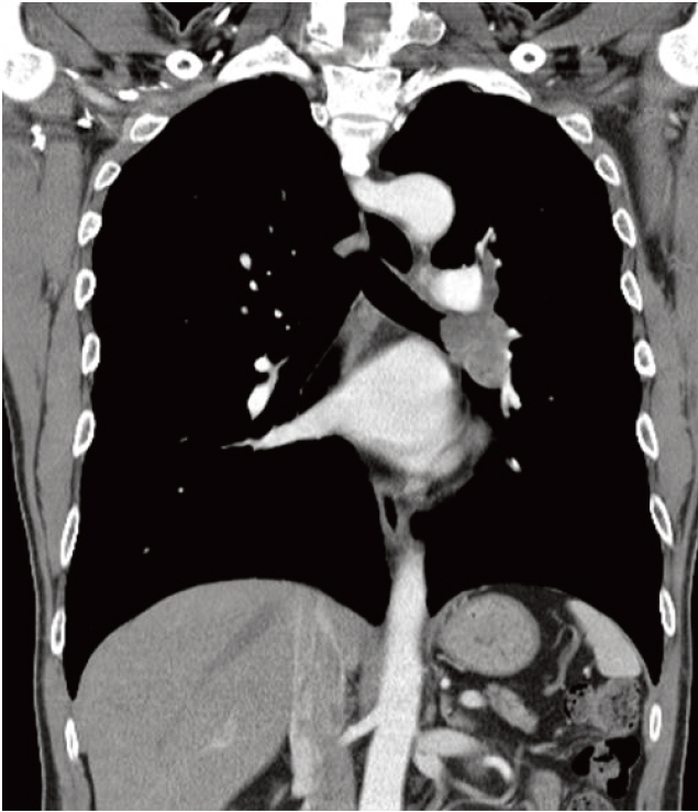
Fig. 3. On the computed tomography images, a mildly enhanced lesion with finger-like appearance was noted along the left main bronchus extending to the upper and lower lobar bronchi, as suspected on digital tomosynthesis.
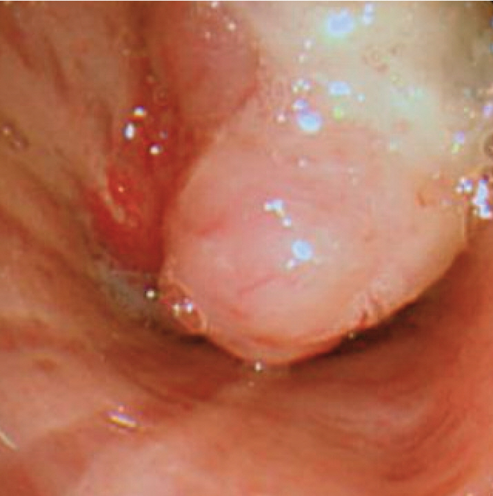
Fig. 4. Bronchoscopic finding. Subsequent bronchoscopy revealed a mass lesion almost completely occluding the left main bronchus at a site 4 cm below the carina and protruding to the endobronchial space. Bronchoscopic biopsy was performed.
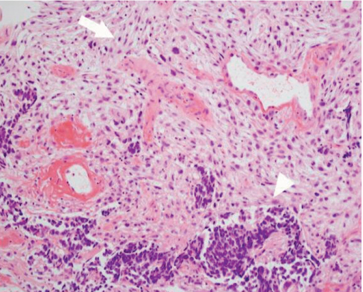
Fig. 5. Pathologic finding demonstrated sarcomatous (arrow) and carcinomatous (arrowhead) components (H&E staining, ×200).
Cited by 1 articles
-
A Rare Case of Tracheal Leiomyoma: Role of Digital Tomosynthesis in Diagnosis and Treatment
Soo Won Nam, Yeon Joo Jeong, Geewon Lee, Ji Won Lee, Jung Seop Eom, Chang Hun Lee, So Min Park
J Korean Soc Radiol. 2020;81(1):225-230. doi: 10.3348/jksr.2020.81.1.225.
Reference
-
References
1. Huwer H, Kalweit G, Straub U, Feindt P, Volkmer I, Gams E. Pulmonary carcinosarcoma: diagnostic problems and determinants of the prognosis. Eur J Cardiothorac Surg. 1996; 10:403–7.
Article2. Sokucu SN, Kocaturk C, Urer N, Sonmezoglu Y, Dalar L, Karasulu L, et al. Evaluation of six patients with pulmonary carcinosarcoma with a literature review. ScientificWorldJournal. 2012; 2012:167317.3. Chen Q, Goo JM, Seo JB, Chung MJ, Lee YJ, Im JG. Evaluation of tracheobronchial diseases: comparison of different imaging techniques. Korean J Radiol. 2000; 1:135–41.
Article4. Bath M, Svalkvist A, von Wrangel A, Rismyhr-Olsson H, Cederblad A. Effective dose to patients from chest examinations with tomosynthesis. Radiat Prot Dosimetry. 2010; 139:153–8.
Article5. Kim EY, Chung MJ, Lee HY, Koh WJ, Jung HN, Lee KS. Pulmonary mycobacterial disease: diagnostic performance of low-dose digital tomosynthesis as compared with chest radiography. Radiology. 2010; 257:269–77.
Article6. Vult von Steyern K, Bjorkman-Burtscher IM, Weber L, Hoglund P, Geijer M. Effective dose from chest tomosynthesis in children. Radiat Prot Dosimetry. 2014; 158:290–8.
Article7. Vikgren J, Zachrisson S, Svalkvist A, Johnsson AA, Boijsen M, Flinck A, et al. Comparison of chest tomosynthesis and chest radiography for detection of pulmonary nodules: human observer study of clinical cases. Radiology. 2008; 249:1034–41.
Article8. Kim KI, Flint JD, Muller NL. Pulmonary carcinosarcoma: radiologic and pathologic findings in three patients. AJR Am J Roentgenol. 1997; 169:691–4.
Article9. Yoshino N, Kubokura H, Yamauchi S, Ohaki Y, Koizumi K, Shimizu K. A true pulmonary carcinosarcoma that required diagnostic differentiation from a pleomorphic adenoma: a case report. Ann Thorac Cardiovasc Surg. 2009; 15:42–5.10. Zehani A, Ayadi-Kaddour A, Mlika M, Hamrouni R, Fkih L, Marghli A, et al. Primary pulmonary carcinosarcoma. Tunis Med. 2013; 91:287–9.
- Full Text Links
-
- Actions
-
Cited
- CITED
-
- Close
- Share
-
- Similar articles
-
- Airway obstruction by dislodgement of an endobronchial tumor fragment during right lung lobectomy using a bronchial blocker: A case report
- Airway obstruction in heat & moisture exchanger filter: A case report
- Comparative Study between Digital Tomosynthesis and Endoscopic Retrograde Cholangiopancreatography for the Evaluation of Common Bile Duct Stones: Focus on Detection and Stone Conspicuity
- Clinical Application of Artificial Intelligence in Digital Breast Tomosynthesis
- A Case of Tracheal Tumor Presented as Bronchial AsthmaA Case of Tracheal Tumor Presented as Bronchial Asthma
