The Comparative Morphometric Study of the Posterior Cranial Fossa : What Is Effective Approaches to the Treatment of Chiari Malformation Type 1?
- Affiliations
-
- 1Department of Neurosurgery, Dongtan Sacred Heart Hospital, College of Medicine, Hallym University, Hwaseong, Korea.
- 2Department of Neurosurgery, Kangnam Sacred Heart Hospital, College of Medicine, Hallym University, Seoul, Korea.
- 3Department of Neurosurgery, Kangdong Sacred Heart Hospital, College of Medicine, Hallym University, Seoul, Korea.
- 4Department of Neurosurgery, Sacred Heart Hospital, College of Medicine, Hallym University, Anyang, Korea.
- 5Department of Neurosurgery, Soonchunhyang Unviersity Bucheon Hospital, Bucheon, Korea. neuri71@gmail.com
- KMID: 2138356
- DOI: http://doi.org/10.3340/jkns.2013.54.5.405
Abstract
OBJECTIVE
The objective of this study was to investigate changes in the posterior cranial fossa in patients with symptomatic Chiari malformation type I (CMI) compared to a control group.
METHODS
We retrospectively reviewed clinical and radiological data from 12 symptomatic patients with CMI and 24 healthy control subjects. The structures of the brain and skull base were investigated using magnetic resonance imaging.
RESULTS
The length of the clivus had significantly decreased in the CMI group than in the control group (p=0.000). The angle between the clivus and the McRae line (p<0.024), as the angle between the supraocciput and the McRae line (p<0.021), and the angle between the tentorium and a line connecting the internal occipital protuberance to the opisthion (p<0.009) were significantly larger in the CMI group than in the control group. The mean vertical length of the cerebellar hemisphere (p<0.003) and the mean length of the coronal and sagittal superoinferior aspects of the cerebellum (p<0.05) were longer in the CMI group than in the control group, while the mean length of the axial anteroposterior aspect of the cerebellum (p<0.001) was significantly shorter in the CMI group relative to control subjects.
CONCLUSION
We elucidate the transformation of the posterior cranial fossa into the narrow funnel shape. The sufficient cephalocaudal extension of the craniectomy of the posterior cranial fossa has more decompression effect than other type extension of the craniectomy in CMI patients.
Keyword
MeSH Terms
Figure
-
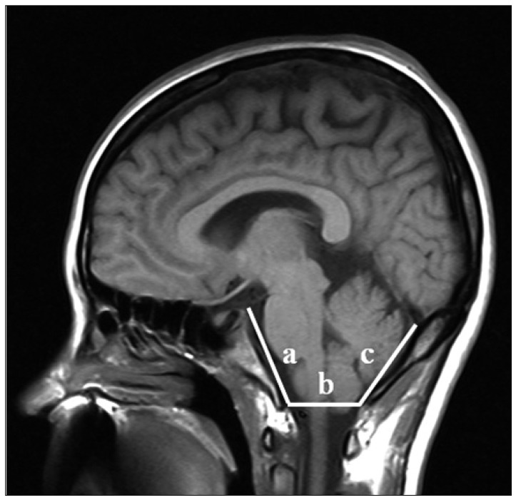
Fig. 1 Midsagittal T1-weighted MRI shows the posterior cranial fossa, the brainstem and cerebellum. a : length of clivus, b : the anteroposterior length of the foramen magnum from distance between the basion and opisthion on the McRae line, c : the length of the supraocciput between the internal occipital protuberance and the opisthion.
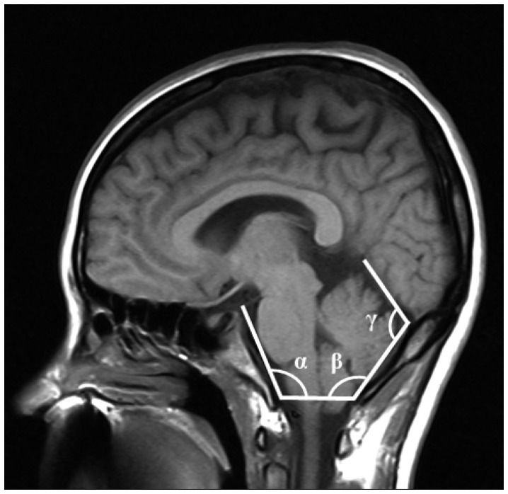
Fig. 2 Midsagittal T1-weighted MRI illustrates demonstrating the measurements of three structural angulations. α : angle of clivus, β : angle of the cerebellar tentorium, γ : angle of occipital protuberance.
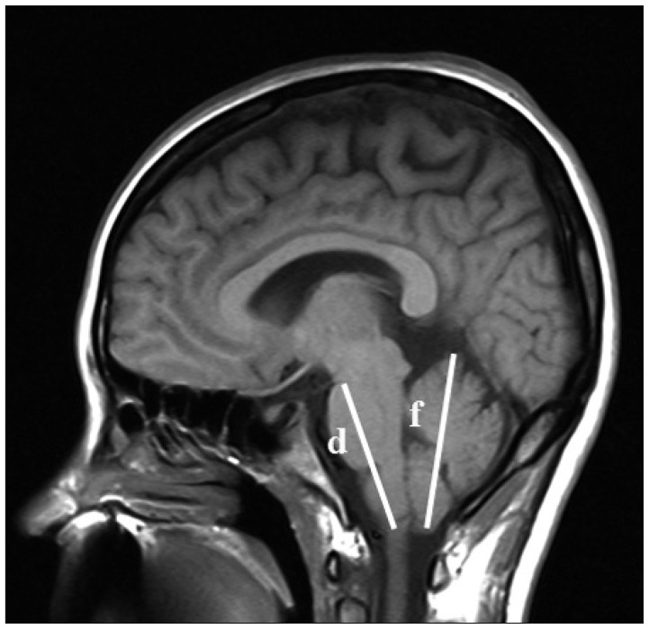
Fig. 3 Midsagittal T1-weighted MRI shows the neural structure of the posterior cranial fossa. d : the length of the hindbrain midbrain-pons junction and the medullocervical junction, f : the vertical length of the tentorium with cerebellum.
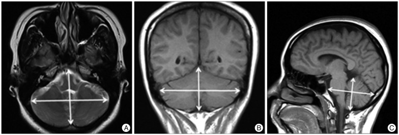
Fig. 4 Demonstration of linear measurements for the maxium diameters of posterior fossa on T2-weighted MRI. (A) Anteroposterior length and bi-cerebellar length of cerebellum on axial view, (B) supero-inferior length and bi-cerebellar diameter of cerebellum on coronal view, (C) supero-inferior length and anteroposterior length of cerebellum on sagittal view.
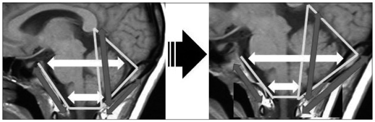
Fig. 5 Schematic illustration of the changes of the posterior cranial fossa in the Chiari malformation type I (CMI) patinets.
Reference
-
1. Aydin S, Hanimoglu H, Tanriverdi T, Yentur E, Kaynar MY. Chiari type I malformations in adults : a morphometric analysis of the posterior cranial fossa. Surg Neurol. 2005; 64:237–241. discussion 241. PMID: 16099255.
Article2. Bindal AK, Dunsker SB, Tew JM Jr. Chiari I malformation: classification and management. Neurosurgery. 1995; 37:1069–1074. PMID: 8584146.3. Blagodatsky MD, Larionov SN, Alexandrov YA, Velm AI. Surgical treatment of Chiari I malformation with or without syringomyelia. Acta Neurochir (Wien). 1999; 141:963–968. PMID: 10526077.
Article4. Cristante L, Westphal M, Herrmann HD. Cranio-cervical decompression for Chiari I malformation. A retrospective evaluation of functional outcome with particular attention to the motor deficits. Acta Neurochir (Wien). 1994; 130:94–100. PMID: 7725949.5. Dagtekin A, Avci E, Kara E, Uzmansel D, Dagtekin O, Koseoglu A, et al. Posterior cranial fossa morphometry in symptomatic adult Chiari I malformation patients: comparative clinical and anatomical study. Clin Neurol Neurosurg. 2011; 113:399–403. PMID: 21333437.
Article6. Dyste GN, Menezes AH, VanGilder JC. Symptomatic Chiari malformations. An analysis of presentation, management, and long-term outcome. J Neurosurg. 1989; 71:159–168. PMID: 2746341.7. Ergün R, Akdemir G, Gezici AR, Tezel K, Beskonakli E, Ergüngör F, et al. Surgical management of syringomyelia-Chiari complex. Eur Spine J. 2000; 9:553–557. PMID: 11189926.
Article8. Fischer EG. Posterior fossa decompression for Chiari I deformity, including resection of the cerebellar tonsils. Childs Nerv Syst. 1995; 11:625–629. PMID: 8608577.
Article9. Fujii K, Natori Y, Nakagaki H, Fukui M. Management of syringomyelia associated with Chiari malformation : comparative study of syrinx size and symptoms by magnetic resonance imaging. Surg Neurol. 1991; 36:281–285. PMID: 1948628.
Article10. Gambardella G, Caruso G, Caffo M, Germanò A, La Rosa G, Tomasello F. Transverse microincisions of the outer layer of the dura mater combined with foramen magnum decompression as treatment for syringomyelia with Chiari I malformation. Acta Neurochir (Wien). 1998; 140:134–139. PMID: 10398992.
Article11. Guyotat J, Bret P, Jouanneau E, Ricci AC, Lapras C. Syringomyelia associated with type I Chiari malformation. A 21-year retrospective study on 75 cases treated by foramen magnum decompression with a special emphasis on the value of tonsils resection. Acta Neurochir (Wien). 1998; 140:745–754. PMID: 9810440.
Article12. Heiss JD, Patronas N, DeVroom HL, Shawker T, Ennis R, Kammerer W, et al. Elucidating the pathophysiology of syringomyelia. J Neurosurg. 1999; 91:553–562. PMID: 10507374.
Article13. Hida K, Iwasaki Y, Koyanagi I, Sawamura Y, Abe H. Surgical indication and results of foramen magnum decompression versus syringosubarachnoid shunting for syringomyelia associated with Chiari I malformation. Neurosurgery. 1995; 37:673–678. discussion 678-679. PMID: 8559295.
Article14. Isu T, Sasaki H, Takamura H, Kobayashi N. Foramen magnum decompression with removal of the outer layer of the dura as treatment for syringomyelia occurring with Chiari I malformation. Neurosurgery. 1993; 33:844–849. discussion 849-850. PMID: 8264881.
Article15. James HE, Brant A. Treatment of the Chiari malformation with bone decompression without durotomy in children and young adults. Childs Nerv Syst. 2002; 18:202–206. PMID: 12042917.
Article16. Klekamp J, Batzdorf U, Samii M, Bothe HW. The surgical treatment of Chiari I malformation. Acta Neurochir (Wien). 1996; 138:788–801. PMID: 8869706.
Article17. McGirt MJ, Nimjee SM, Floyd J, Bulsara KR, George TM. Correlation of cerebrospinal fluid flow dynamics and headache in Chiari I malformation. Neurosurgery. 2005; 56:716–721. discussion 716-721. PMID: 15792510.
Article18. McGirt MJ, Nimjee SM, Fuchs HE, George TM. Relationship of cine phase-contrast magnetic resonance imaging with outcome after decompression for Chiari I malformations. Neurosurgery. 2006; 59:140–146. discussion 140-146. PMID: 16823310.
Article19. Menezes AH. Craniocervical developmental anatomy and its implications. Childs Nerv Syst. 2008; 24:1109–1122. PMID: 18401563.
Article20. Milhorat TH, Bolognese PA. Tailored operative technique for Chiari type I malformation using intraoperative color Doppler ultrasonography. Neurosurgery. 2003; 53:899–905. discussion 905-906. PMID: 14519223.
Article21. Munshi I, Frim D, Stine-Reyes R, Weir BK, Hekmatpanah J, Brown F. Effects of posterior fossa decompression with and without duraplasty on Chiari malformation-associated hydromyelia. Neurosurgery. 2000; 46:1384–1389. discussion 1389-1390. PMID: 10834643.
Article22. Nishikawa M, Sakamoto H, Hakuba A, Nakanishi N, Inoue Y. Pathogenesis of Chiari malformation: a morphometric study of the posterior cranial fossa. J Neurosurg. 1997; 86:40–47. PMID: 8988080.
Article23. Noudel R, Jovenin N, Eap C, Scherpereel B, Pierot L, Rousseaux P. Incidence of basioccipital hypoplasia in Chiari malformation type I : comparative morphometric study of the posterior cranial fossa. Clinical article. J Neurosurg. 2009; 111:1046–1052. PMID: 19463049.
Article24. Raftopoulos C, Sanchez A, Matos C, Balériaux D, Bank WO, Brotchi J. Hydrosyringomyelia-Chiari I complex. Prospective evaluation of a modified foramen magnum decompression procedure: preliminary results. Surg Neurol. 1993; 39:163–169. PMID: 8351630.
Article25. Sekula RF Jr, Jannetta PJ, Casey KF, Marchan EM, Sekula LK, McCrady CS. Dimensions of the posterior fossa in patients symptomatic for Chiari I malformation but without cerebellar tonsillar descent. Cerebrospinal Fluid Res. 2005; 2:11. PMID: 16359556.
Article26. Sindou M, Chávez-Machuca J, Hashish H. Cranio-cervical decompression for Chiari type I-malformation, adding extreme lateral foramen magnum opening and expansile duroplasty with arachnoid preservation. Technique and long-term functional results in 44 consecutive adult cases -- comparison with literature data. Acta Neurochir (Wien). 2002; 144:1005–1019. PMID: 12382129.
Article27. Trigylidas T, Baronia B, Vassilyadi M, Ventureyra EC. Posterior fossa dimension and volume estimates in pediatric patients with Chiari I malformations. Childs Nerv Syst. 2008; 24:329–336. PMID: 17657497.
Article28. Tsara V, Serasli E, Kimiskidis V, Papagianopoulos S, Katsaridis V, Fylaktakis M, et al. Acute respiratory failure and sleep-disordered breathing in Arnold-Chiari malformation. Clin Neurol Neurosurg. 2005; 107:521–524. PMID: 16202827.
Article
- Full Text Links
-
- Actions
-
Cited
- CITED
-
- Close
- Share
-
- Similar articles
-
- Whole-spinal Syringomyelia in a Pediatric Patient with Chiari Malformation Type 1
- Cerebellar Ectopia Associated with Unilateral Agenesis of Posterior Arch of Atlas
- Experimentally induced Chiari-like malformation with myeloschisis in chick embryos
- Posterior Fossa Decompression in Syringomyelia with Chiari Malformation
- Acquired Chiari-malformation after Ventriculoperitoneal Shunt for Hydrocepalus Associated with Neurocysticercosis
