Guide bone regeneration using autogenous teeth: case reports
- Affiliations
-
- 1Department of Oral and Maxillofacial Surgery, Section of Dentistry, Seoul National University Bundang Hospital, Seongnam, Korea.
- 2Department of Periodontology, Section of Dentistry, Seoul National University Bundang Hospital, Seongnam, Korea.
- 3Department of Oral and Maxillofacial Surgery, College of Dentistry, Dankook University, Cheonan, Korea.
- 4Department of Oral and Maxillofacial Surgery, School of Dentistry, Chosun University, Gwangju, Korea.
- 5Research & Development Center, Korea Auto-teeth & Bank, Seoul, Korea. kyk0505@snubh.org
- KMID: 2137005
- DOI: http://doi.org/10.5125/jkaoms.2011.37.2.142
Abstract
- The authors installed implants combined with guided bony regeneration (GBR) using autogenous tooth bone graft material in the patients. In one patient, GBR and simultaneous implant placement were performed. In two patients, GBR was performed and the implants were placed after 6 months. All patients achieved favorable clinical outcomes. Excellent osteoconductive bony healing was observed in the 6 month histology examination after the bone graft.
Figure
-

Fig. 1. Initial panoramic radiography.
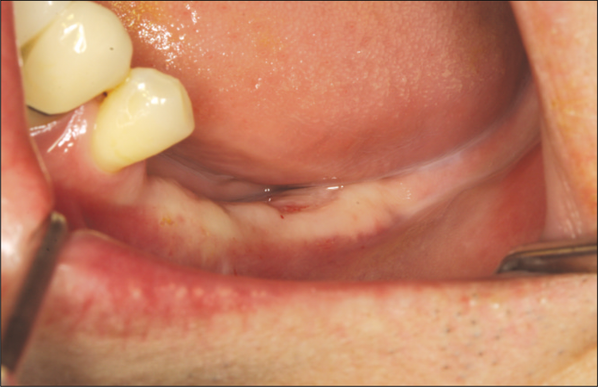
Fig. 2. Preoperative intraoral view. Teeth were extracted 2 months ago.
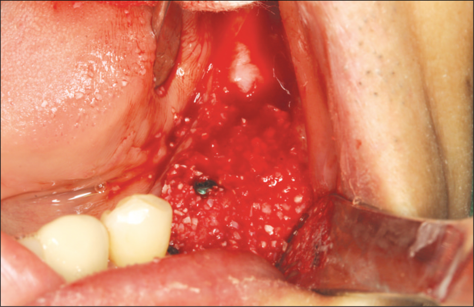
Fig. 3. Implants were placed and dehiscence defects were covered autogenous tooth bone graft material.
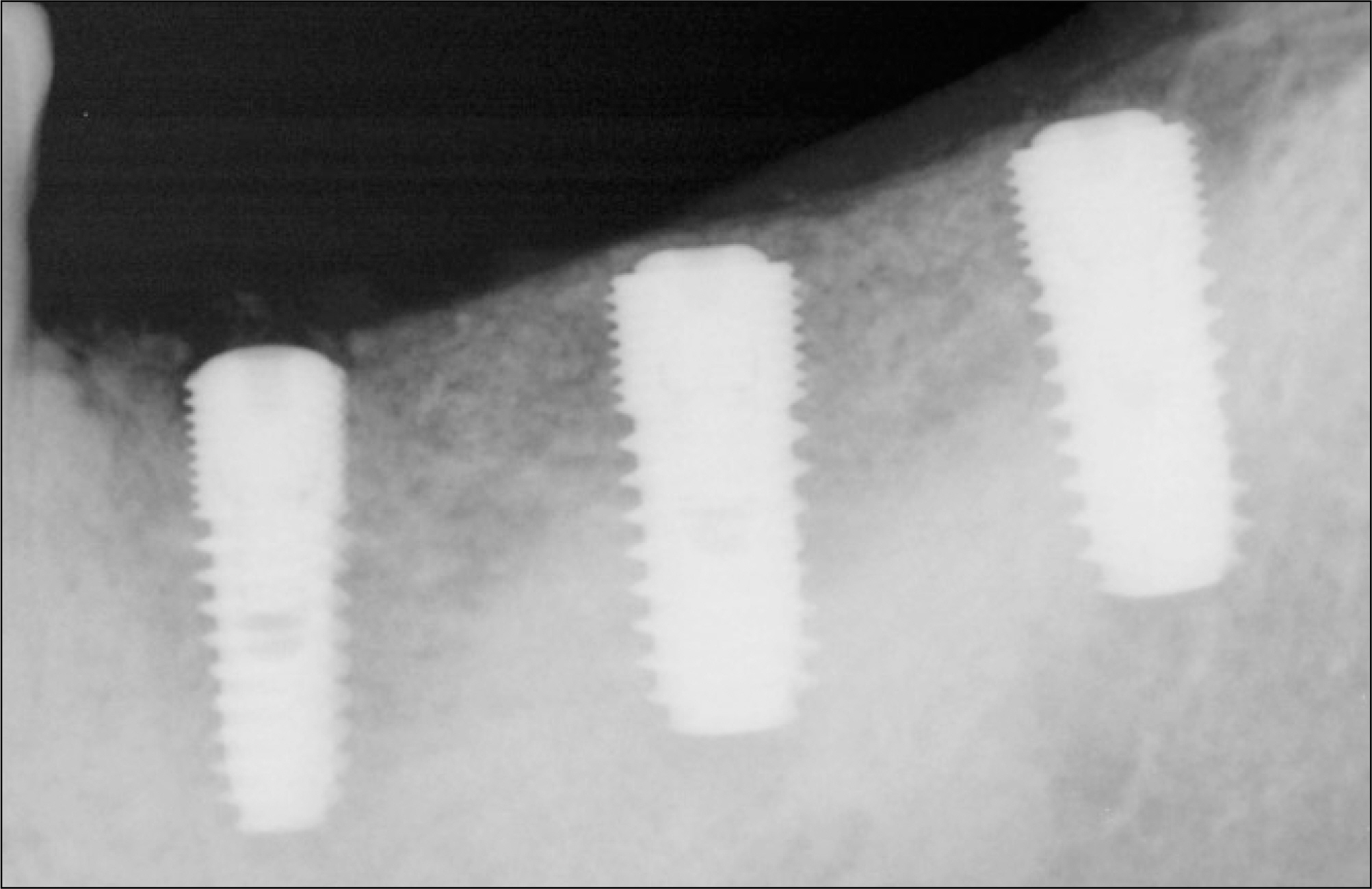
Fig. 4. Periapical radiography 6 months after implant placement.

Fig. 5. Secondary surgery was performed and flap was elevated. Excellent bony healing was observed.
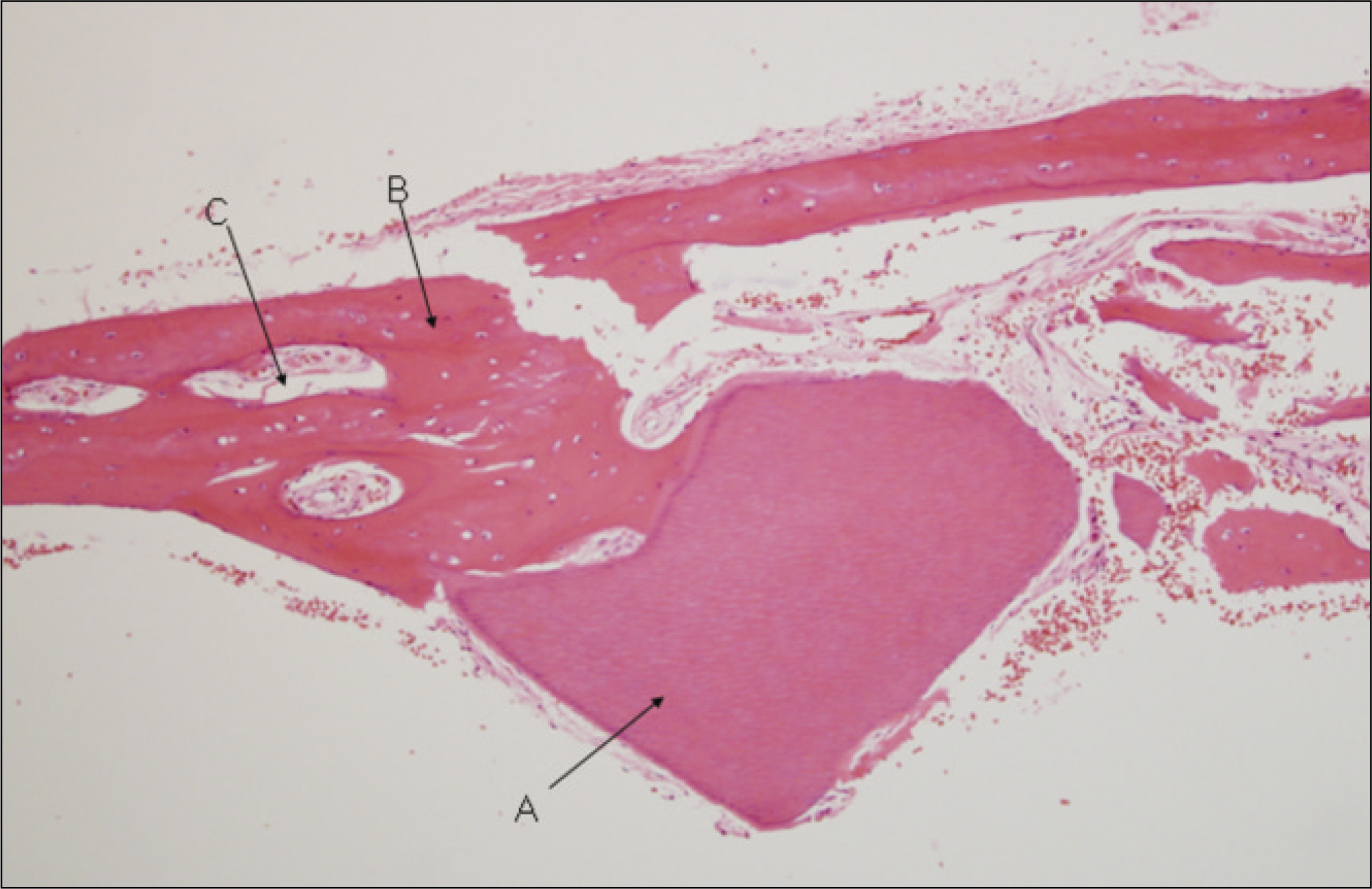
Fig. 6. The remodeling of new bones formed in the vicinity of graft materials is observed. A was graft materials, B was newly-formed bones, C was bone marrow.(H&E staining, original magnification x100)
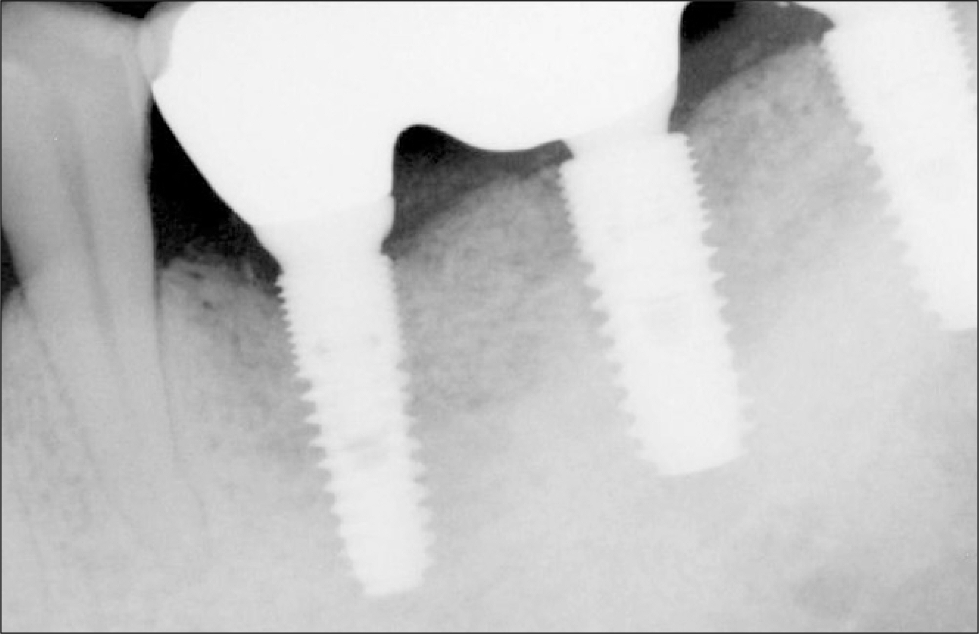
Fig. 7. Periapical radiography 6 months after final prosthetic delivery.
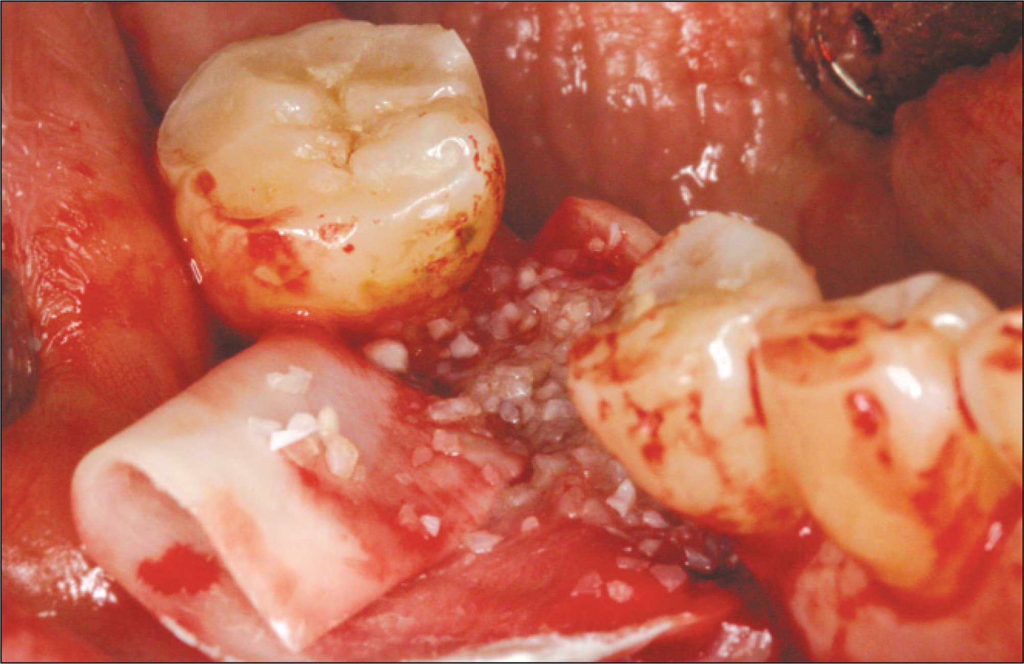
Fig. 8. Guided bony regeneration (GBR) was performed at right 1st molar area of 49-year old female patient. Autogenous tooth bone graft material and collagen membrane were used.
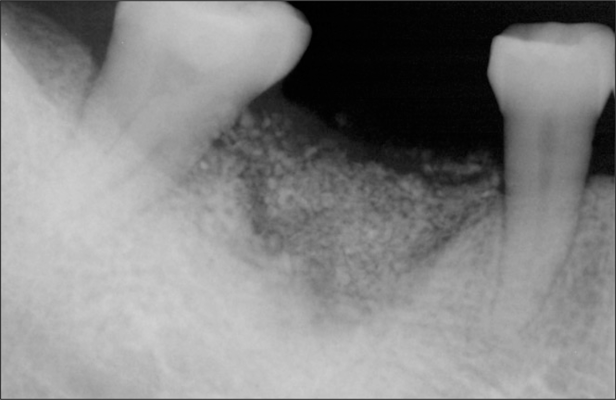
Fig. 9. Periapical radiography 3 weeks after bone graft.

Fig. 10. Periapical radiography 6 months after bone graft. Alveolar crestal level was stable.
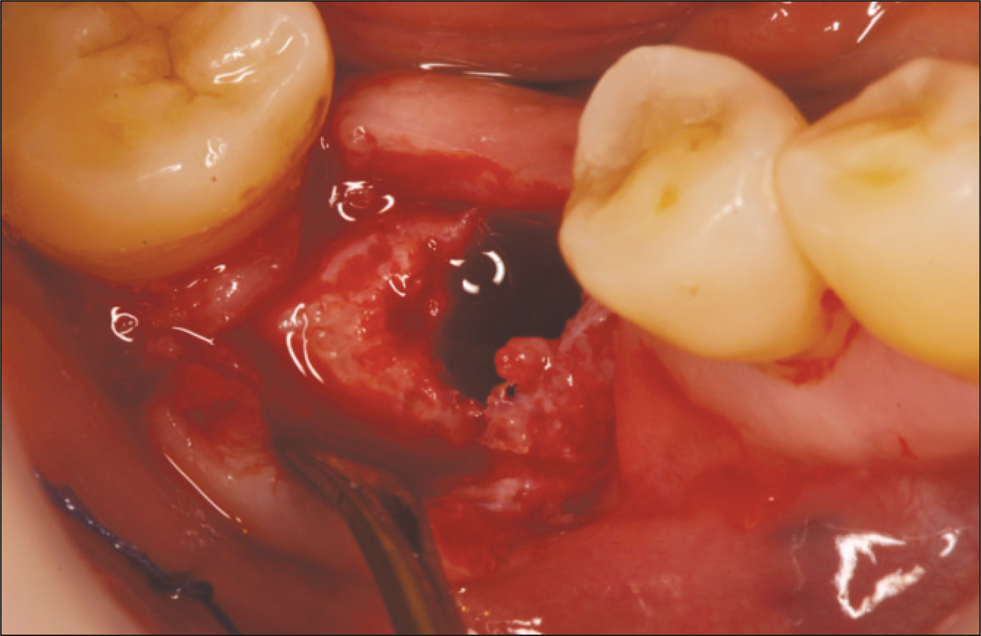
Fig. 11. Implant was installed 6 months after bone graft. Bony quality was type I.
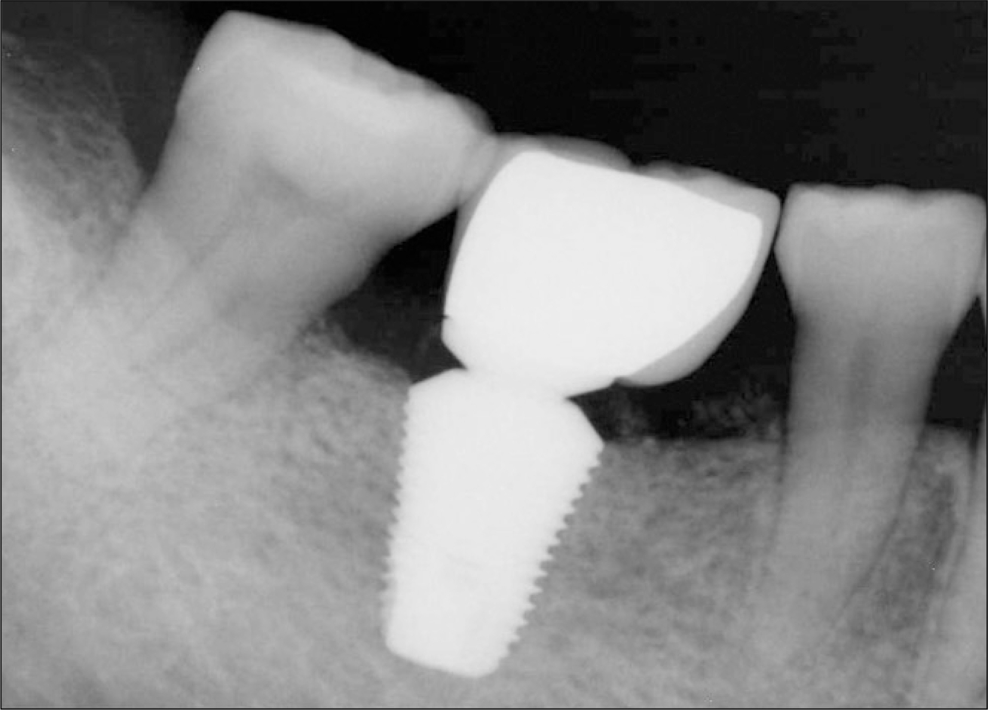
Fig. 12. Periapical radiography after final prosthetic delivery.
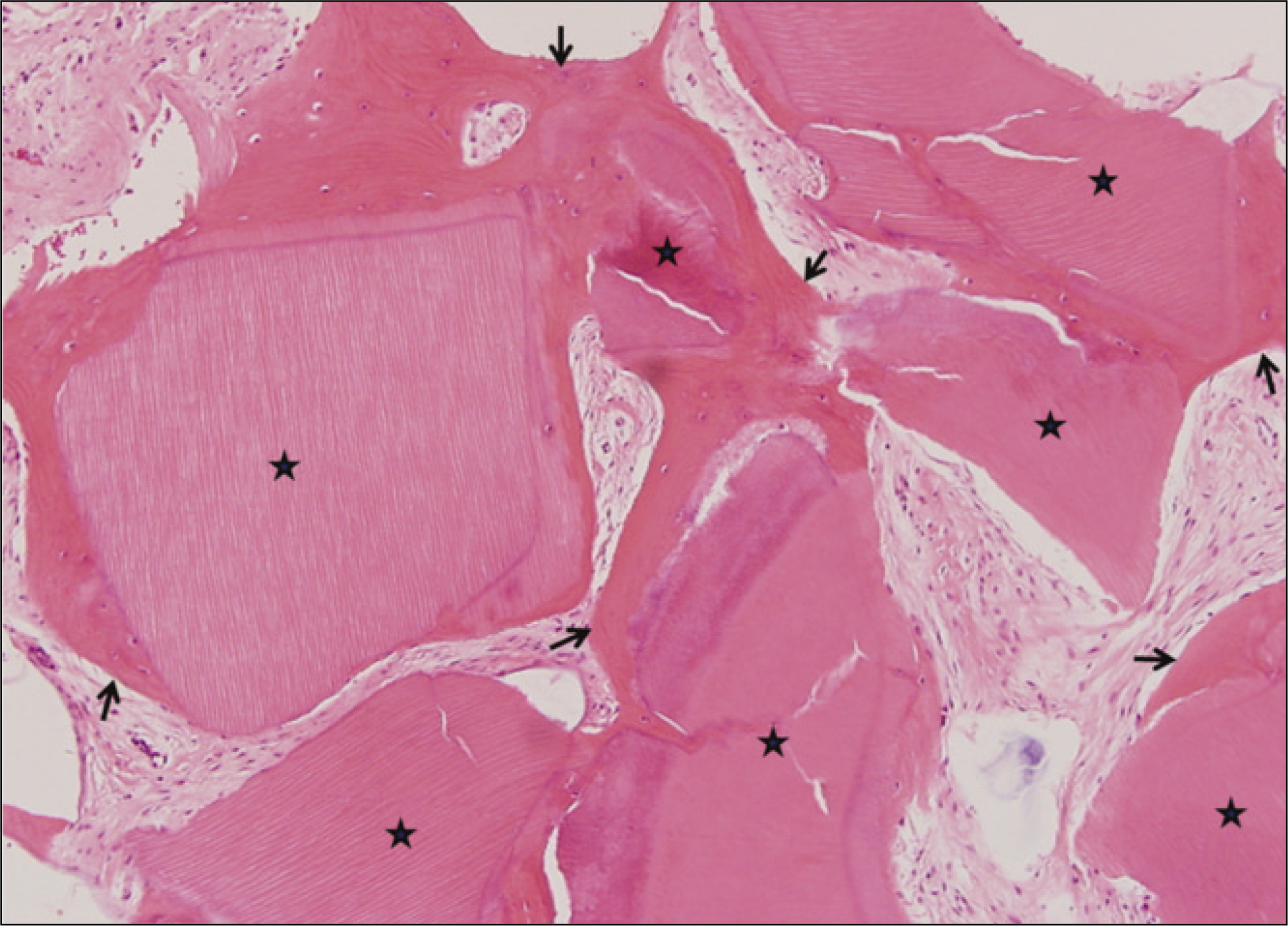
Fig. 13. Microphotograph 6 months after AutoBT transplantation. Higher magnification demonstrated new bone formation (arrows) around the implant chips (asterisks).(H&E staining, original magnification x100)

Fig. 14. Periapical radiography of 50-year old male patient 2 months after extraction of mandibular left 1st molar.

Fig. 15. Periapical radiography 2 weeks after autogenous tooth bone graft.
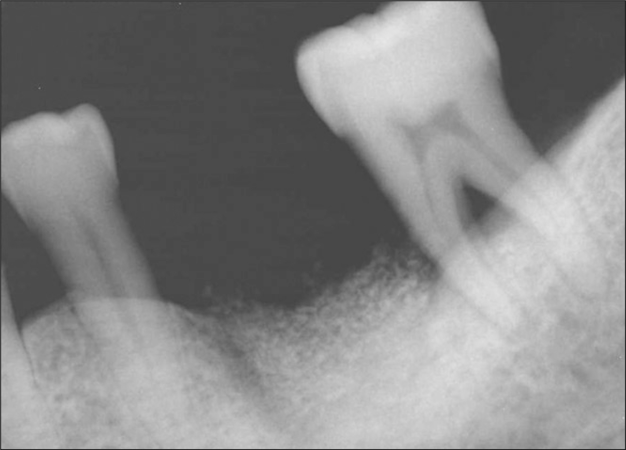
Fig. 16. Periapical radiography 5 months after autogenous tooth bone graft. Alveolar crestal bone level was stable.

Fig. 17. Implant was placed 6 months after bone graft. Adjacent 2nd molar was extracted.
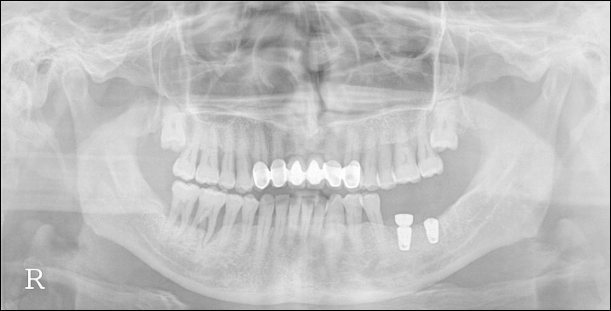
Fig. 18. Second surgery was performed at left 1st molar area. Additional implant was placed at left 2nd molar area.
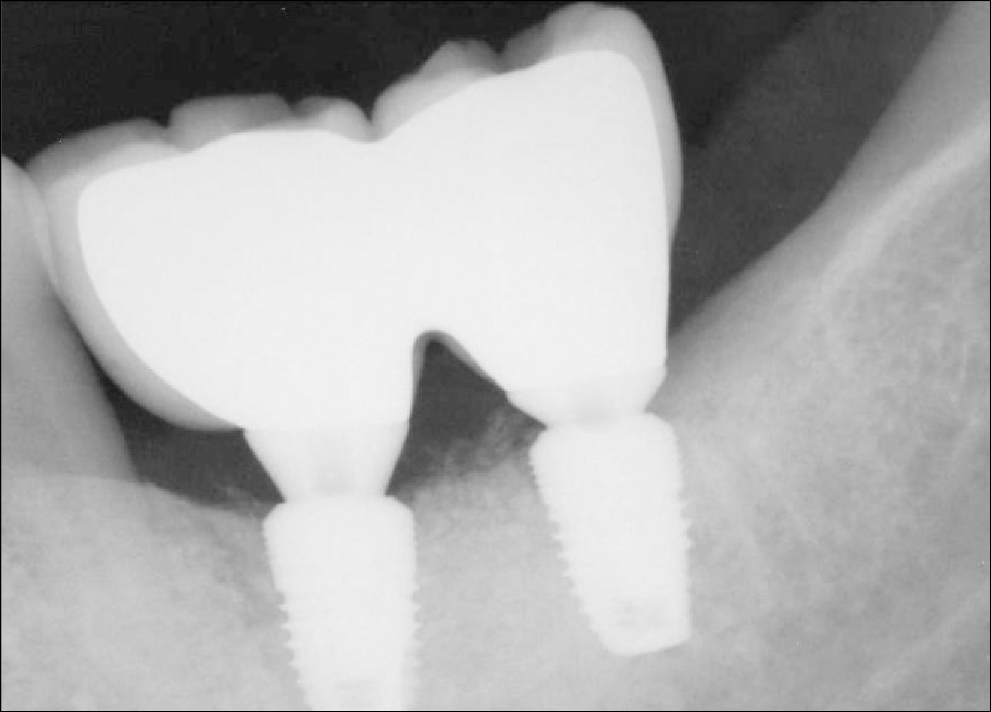
Fig. 19. Periapical radiography after final prosthetic delivery.
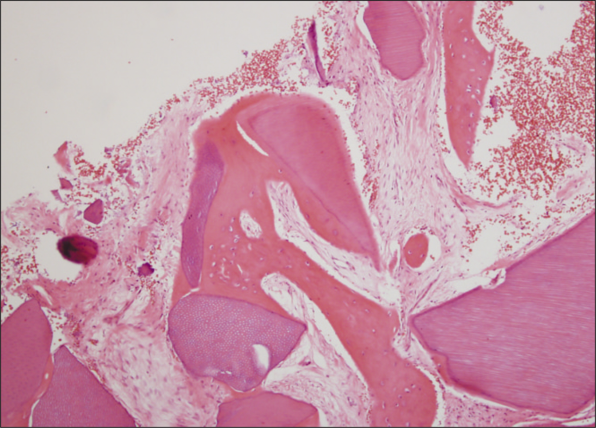
Fig. 20. Microphotograph 6 months after AutoBT transplantation. Higher magnification demonstrated new bone formation around the implant chips.(H&E staining, original magnification x200)
Cited by 4 articles
-
Bone graft material using teeth
Young-Kyun Kim
J Korean Assoc Oral Maxillofac Surg. 2012;38(3):134-138. doi: 10.5125/jkaoms.2012.38.3.134.Comparison of autogenous tooth bone graft and synthetic bone graft materials used for bone resorption around implants after crestal approach sinus lifting: a retrospective study
Young-Kyun Kim, Junho Lee, Ji-Young Yun, Pil-Young Yun, In-Woong Um
J Periodontal Implant Sci. 2014;44(5):216-221. doi: 10.5051/jpis.2014.44.5.216.Tooth-derived bone graft material
Young-Kyun Kim, Junho Lee, In-Woong Um, Kyung-Wook Kim, Masaru Murata, Toshiyuki Akazawa, Masaharu Mitsugi
J Korean Assoc Oral Maxillofac Surg. 2013;39(3):103-111. doi: 10.5125/jkaoms.2013.39.3.103.Clinical evaluation of ridge augmentation using autogenous tooth bone graft material: case series study
Ji-Young Lee, Young-Kyun Kim, Yang-Jin Yi, Joon-Ho Choi
J Korean Assoc Oral Maxillofac Surg. 2013;39(4):156-160. doi: 10.5125/jkaoms.2013.39.4.156.
Reference
-
References
1. Kim MJ, Kim YK, Kim SG. A variety of biomaterial used in dental surgery. Seoul: Narae Publishing Co.;2004.2. Kim YK. inventor;. Tooth plaster and manufacturing method thereof. Korean patent 1019980008980. 1998 Mar 17.3. Kim SG, Kim YK. inventors;. Restorative and grafting material for hard tissue defects prepared from animal teeth. US patent 20030717801. 2003 Nov 19.4. Kim YK, Yeo HH, Ryu CH, Lee HB, Byun UR, Cho JO. An experimental study on the tissue reaction of toothash implanted in mandible body of the mature dog. J Korean Assoc Maxillofac Plast Reconstr Surg. 1993; 15:129–36.5. Kim SG, Yeo HH, Kim YK. Grafting of large defects of the jaws with a particulate dentin-plaster of paris combination. Oral Surg Oral Med Oral Pathol Oral Radiol Endod. 1999; 88:22–5.6. Kim SG, Chung CH, Kim YK, Park JC, Lim SC. Use of particulate dentin-plaster of Paris combination with/without platelet-rich plasma in the treatment of bone defects around implants. Int J Oral Maxillofac Implants. 2002; 17:86–94.7. Kim SY, Kim SG, Lim SC, Bae CS. Effects on bone formation in ovariectomized rats after implantation of tooth ash and plaster of Paris mixture. J Oral Maxillofac Surg. 2004; 62:852–7.
Article8. Kim YK, Kim SG, Byeon JH, Lee HJ, Um IU, Lim SC. Development of a novel bone grafting material using autogenous teeth. Oral Surg Oral Med Oral Pathol Oral Radiol Endod. 2010; 109:496–503.
Article9. Min BM. Oral biochemistry. Seoul: Daehan Narae Publishing, Inc.;2007.10. Bhaskar SN. Orban's Oral histology and embryology. 9th ed.St. Louis: Mosby Co.;1980.11. Choung PH. inventor; Method for extracting tooth protein from extracted tooth. Korean patent 1020020008789. 2002 Feb 19.12. Choung PH. inventor;. Tooth protein extracted from extracted tooth and method for using the same. Korean patent 1020040051812. 2004 Jul 3.
- Full Text Links
-
- Actions
-
Cited
- CITED
-
- Close
- Share
-
- Similar articles
-
- Histologic observation of regenerated bone in human intraosseous lesion following guided tissue regeneration with calcium carbonate implant and autogenous bone graft
- A Comparative Study of the effects of Autogenous and Xenogenic Bone grafts with PRP(Platelet Rich Plasma) technique on Periodontal Regeneration
- Autogenous fresh demineralized tooth graft prepared at chairside for dental implant
- BONE REGENERATION IN COMPOSITE GRAFT OF FREEZE-DRIEDDEMINERALIZED BONE AND HYDROXYLAPATITE IN RABBIT CRANIAL DEFECTS
- The experimental study of the bone regeneration on beta-TCP in rabbit cranial bone
