Epithelial-myoepithelial carcinoma on the superficial lobe of the parotid gland: a case report
- Affiliations
-
- 1Department of Oral and Maxillofacial Surgery, Department of Dentistry, Dong-A University Medical Center, Busan, Korea. samehope@naver.com
- KMID: 2136988
- DOI: http://doi.org/10.5125/jkaoms.2011.37.6.505
Abstract
- Epithelial-myoepithelial carcinoma (EMC) is a low-grade malignant salivary gland neoplasm that was first described in 1972. EMC occurs in the older age group, there is a female predilection and mainly involves the parotid gland. Most authors recommend superficial parotidectomy as a treatment for low-grade malignant tumor in the superficial lobe of parotid gland. The treatment of epithelial-myoepithelial tumors typically includes surgical excision aimed at achieving a R0 resection. This paper reports a case of EMC of the parotid gland treated only by a conservational surgical excision. The lesion was exposed by the retromandibular approach and detached. After the parotid gland envelop was exposed, the mass was observed and was easy to remove due to capsulation. The preoperative diagnosis was a pleomorphic adenoma on the left parotid gland. The tumor was removed surgically with a conservative extracapsular dissection. The postoperative diagnosis was EMC, so superficial parotidectomy or radiation therapy was considered. Nevertheless, the patient was observed and no additional treatment was attempted because the patient was old and a successfully excision of the tumor had been achieved.
Figure
-

Fig. 1. Facial view illustrating the 3–4 cm swelling on patient's left cheek. Sun-Mi Jin et al: Epithelial-myoepithelial carcinoma on the superficial lobe of the parotid gland: a case report. J Korean Assoc Oral Maxillofac Surg 2011
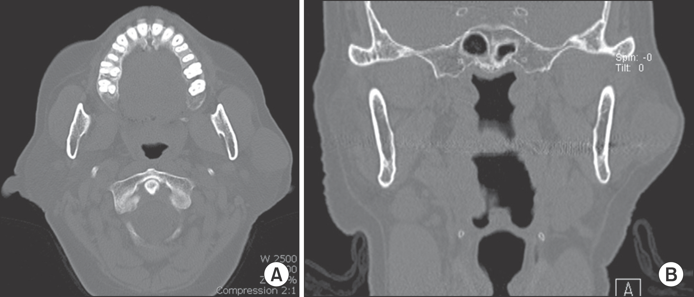
Fig. 2. Computed tomography (CT) scan. A. Transverse view of CT scan. B. Coronal view of CT scan. Sun-Mi Jin et al: Epithelial-myoepithelial carcinoma on the superficial lobe of the parotid gland: a case report. J Korean Assoc Oral Maxillofac Surg 2011
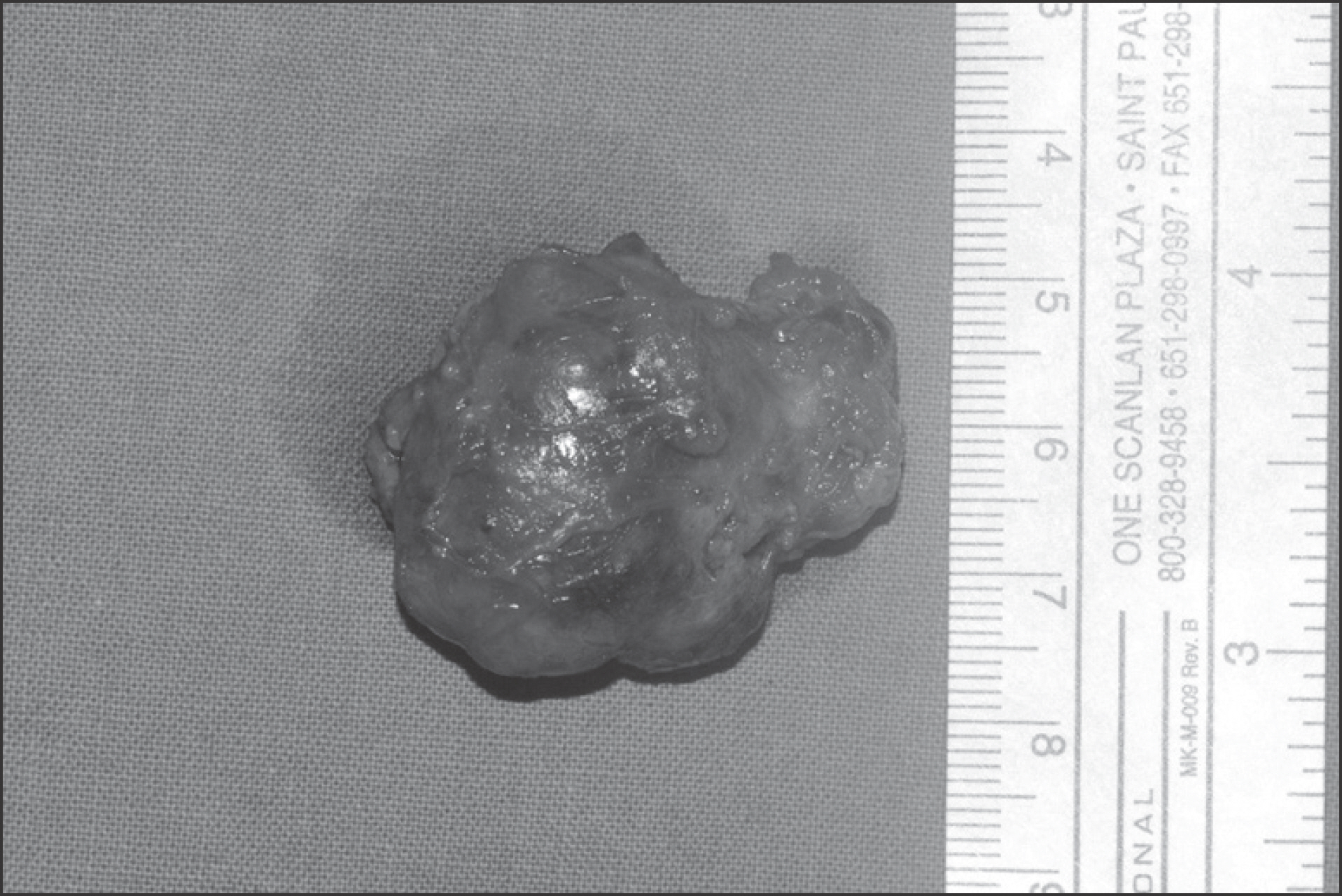
Fig. 3. Macroscopic view (external) shows well-circumscribed, thinly encapsulated mass with soft consistency. Sun-Mi Jin et al: Epithelial-myoepithelial carcinoma on the superficial lobe of the parotid gland: a case report. J Korean Assoc Oral Maxillofac Surg 2011
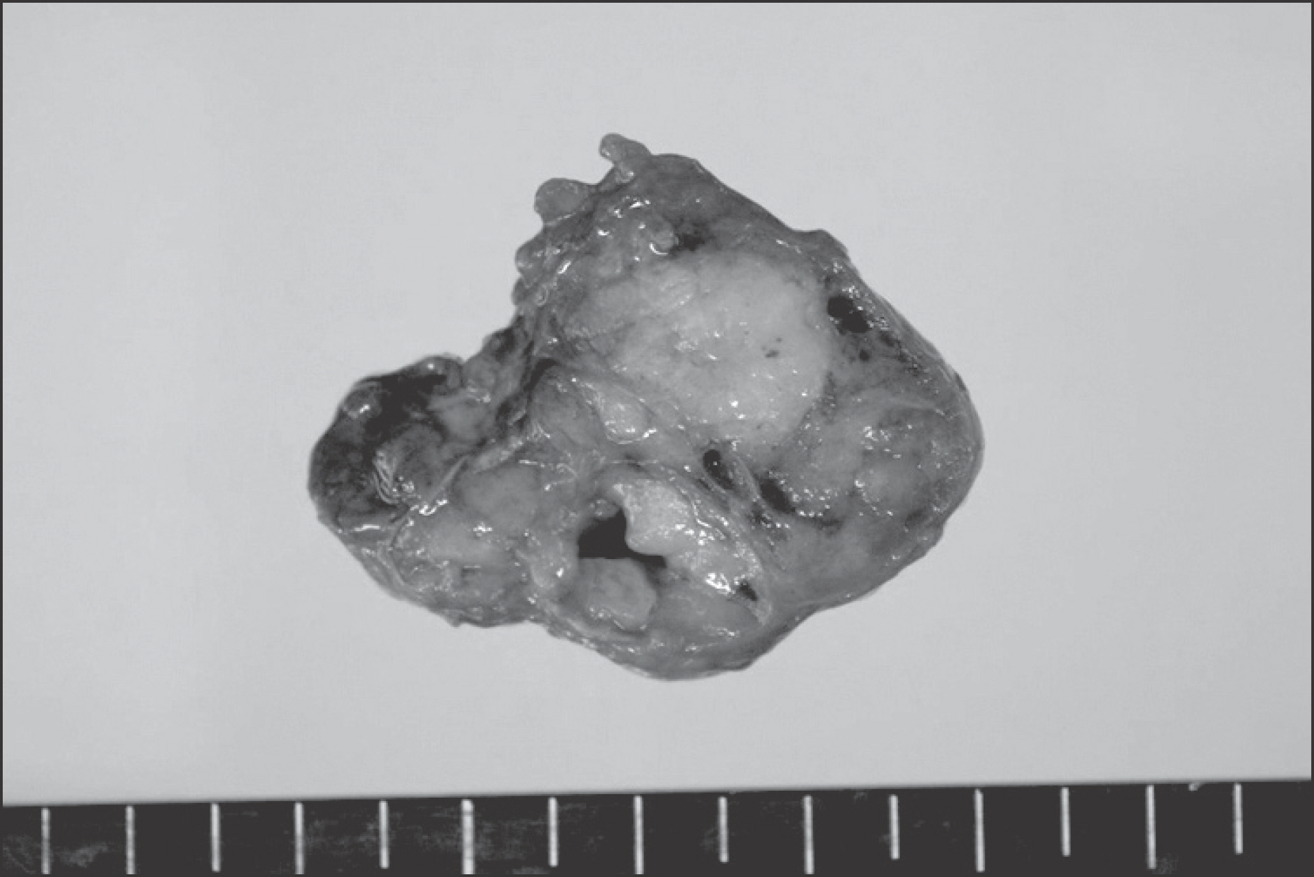
Fig. 4. Macroscopic view (cross section) shows a relatively well defined grayish white soft tissue mass with focal cystic change on sectioning. The tumor is confined to the gland. Sun-Mi Jin et al: Epithelial-myoepithelial carcinoma on the superficial lobe of the parotid gland: a case report. J Korean Assoc Oral Maxillofac Surg 2011
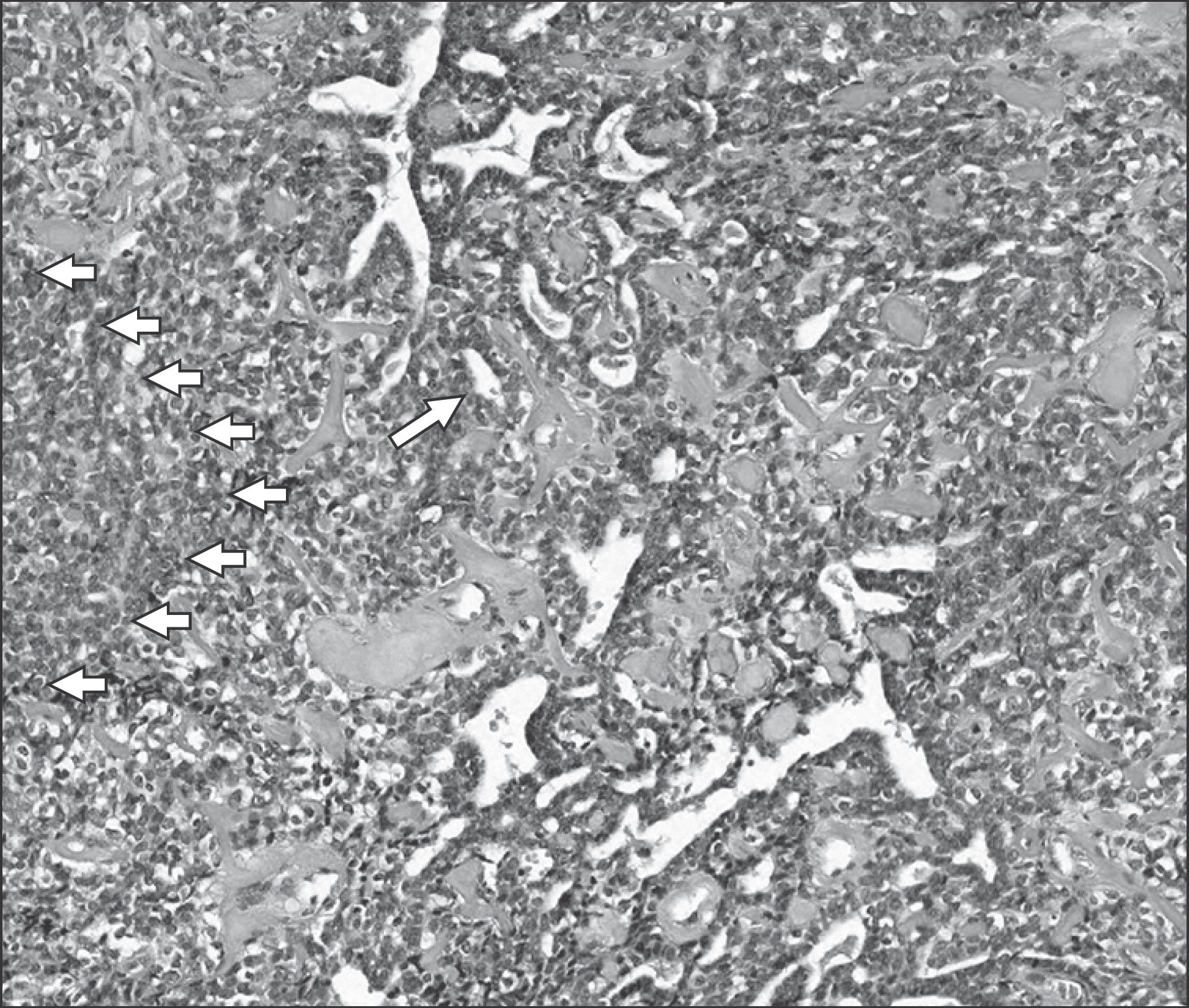
Fig. 5. Microscopic view (H&E staining, ×100). The parotid gland mass is composed of double layered tubules and solid area of clear cells (short arrows). The tubules are lined by inner cuboidal cells with eosinophilic cytoplasm and outer layer of clear, myoepithelial type cells (long arrow). Sun-Mi Jin et al: Epithelial-myoepithelial carcinoma on the superficial lobe of the parotid gland: a case report. J Korean Assoc Oral Maxillofac Surg 2011
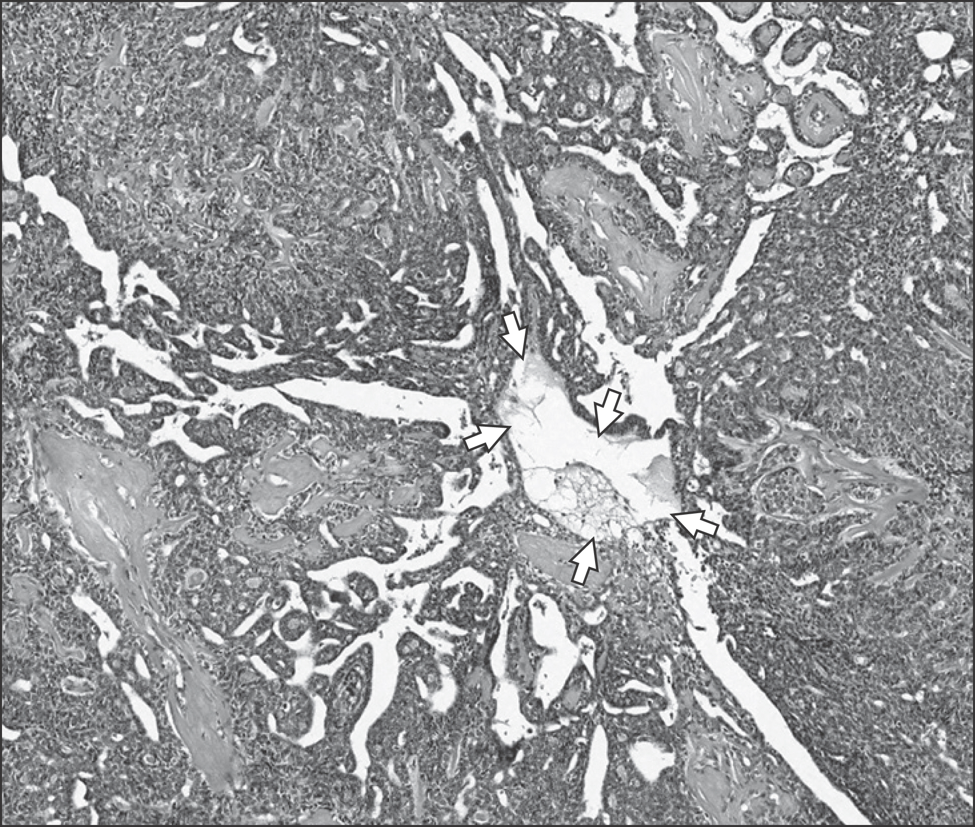
Fig. 6. Microscopic view (H&E staining, ×40). The tumor also shows cystic area with papillary fronds (arrows). Sun-Mi Jin et al: Epithelial-myoepithelial carcinoma on the superficial lobe of the parotid gland: a case report. J Korean Assoc Oral Maxillofac Surg 2011

Fig. 7. The immunohistochemical stain confirms the outer layer of tubules are lined by p63 positive myoepithelial cells (arrows) (anti-p63 immunohistochemistry, ×100). Sun-Mi Jin et al: Epithelial-myoepithelial carcinoma on the superficial lobe of the parotid gland: a case report. J Korean Assoc Oral Maxillofac Surg 2011
Reference
-
References
1. Seifert G, Sobin LH. Epithelial-myoepithelial carcinoma. WHO. World Health Organization International Classification of Tumors: Histological Typing of Salivary Gland Tumors. 2nd ed.Berlin: Spring-Verlag;1991. p. 23–4.2. Dorothy MP. Surgical Pathology and Fine Needle Aspiration Cytopathology. John EN, Mark MG, editors. Color Atlas and Text of the Salivary Glands. 1st ed.Barcelona: Mosby-Wolfe;1995. : 78.3. Suksela E, Tarkkanen J, Wartiovaara J. Parotid clear cell adenoma of possible myoepithelial origin. Cancer. 1972; 30:742–8.4. Bauer WH, Fox RA. Adenomyoepithelioma (Cylindroma) of palatal mucous glands. Arch Pathol. 1945; 39:96–102.5. Donath K, Seifert G, Schmitz R. Diagnosis and ultrastructure of the tubular carcinoma of salivary gland ducts. Epithelial-myoepithelial carcinoma of the intercalated ducts. Virchows Arch. 1972; 356:16–31.6. Yamada H, Kawaguchi K, Yagi M, Morita Y, Mishima K, Uno K, et al. Epithelial-myoepithelial carcinoma of the submandibular gland with a high uptake of 18F-FDG: a case report and image diagnosis. Oral Surg Oral Med Oral Pathol Oral Radiol Endod. 2007; 104:243–8.
Article7. Kokemueller H, Swennen G, Brueggemann N, Brachvogel P, Eckardt A, Hausamen JE. Epithelial malignancies of the salivary glands: clinical experience of a single institution-a review. Int J Oral Maxillofac Surg. 2004; 33:423–32.
Article8. Baek S, Ha JW, Oh HK, Ryu SY, Kim WJ. A clinical study on the parotid gland tumors. J Korean Assoc Maxillofac Plastic Reconstructive Surg. 2002; 24:398–405.9. Woods JE, Chong GC, Beahrs OH. Experience with 1,360 primary parotid tumors. Am J Surg. 1975; 130:460–2.
Article10. Park TW, Lee SL, Kim JD, Park CS, Choi SC, Ko KJ, et al. Oral and Maxillofacial Radiology. 3rd ed.Seoul: Narae Publishing;2001. p. 476–7.11. Colio RL, Sciubba JJ, Brannon RB, Batsakis JG. Epithelial/myoepithelial carcinoma of intercalated duct origin. A clinicopathologic and ultrastructural assessment of sixteen cases. Oral Surg Oral Med Oral Pathol. 1982; 53:280–7.12. Cheung FM, Hioe F, Kong JH. Histologic variant of the epithelial/myoepithelial carcinoma of the salivary gland: a case report. Head Neck. 1995; 17:437–44.
Article13. Di Palna S. Epithelial/myoepithelial carcinoma with coexisting multifocal intercalated duct hyperplasia of the parotid gland. Histopathology. 1994; 25:494–6.
Article14. Deere H, Hore I, McDermott N, Levine T. Epithelial-myoepithelial carcinoma of the parotid gland: a case report and review of the cytological and histological features. J Laryngol Otol. 2001; 115:434–6.
Article15. Spiro RH, Huvos AG, Strong EW. Cancer of the parotid gland. A clinicopathologic study of 288 primary cases. Am J Surg. 1975; 130:452–9.16. Harish K. Management of primary malignant epithelial parotid tumors. Surgical Oncol. 2004; 13:7–16.
Article17. Lim YC, Lee SY, Kim KB, Lee JS, Koo BS, Shin HA, et al. Conservative parotidectomy for the treatment of parotid cancers. Oral Oncol. 2005; 41:1021–7.
Article
- Full Text Links
-
- Actions
-
Cited
- CITED
-
- Close
- Share
-
- Similar articles
-
- A Case of Epithelial-myoepithelial Carcinoma of the Parotid Gland
- Combined Epithelial-Myoepithelial Carcinoma and Basal Cell Adenocarcinoma of the Parotid Gland: A case report
- Epithelial-Myoepithelial Carcinoma of the Lung: one case report
- A Case of Myoepithelial Carcinoma Originated from Inferior Turbinate
- Epithelial-Myoepithelial Carcinoma of Intercalated Duct of Parotid Gland
