J Clin Neurol.
2010 Mar;6(1):41-45. 10.3988/jcn.2010.6.1.41.
Benign Oligemia Despite a Malignant MRI Profile in Acute Ischemic Stroke
- Affiliations
-
- 1Department of Neurology and the Stroke and Cerebrovascular Center, Samsung Medical Center, Sungkyunkwan University School of Medicine, Seoul, Korea. nmboy@unitel.co.kr
- 2Department of Neurology, UCLA Stroke Center, Los Angeles, CA, USA.
- KMID: 2135481
- DOI: http://doi.org/10.3988/jcn.2010.6.1.41
Abstract
- BACKGROUND
It has recently been suggested that diffusion and perfusion MRI can identify subgroups likely to benefit or potentially be harmed by reperfusion therapies.
CASE REPORT
We investigated serial MRI data of two patients with occlusion of the proximal middle cerebral artery (MCA). In both cases, acute multiple cortical infarcts evident on diffusion-weighted imaging (DWI) and perfusion-weighted imaging (PWI) showed extensive areas of severe perfusion delays, indicating a malignant MRI profile. However, despite the malignant MRI profiles in these cases, no new ischemic lesions or hemorrhage evolved even in the presence of persistent arterial occlusion, and the patients recovered without sequelae.
CONCLUSIONS
These two cases suggest that time-domain PWI findings should be interpreted with caution in certain scenarios of acute ischemic stroke.
Keyword
MeSH Terms
Figure
-
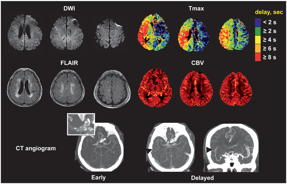
Fig. 1 MRI and CT angiography findings for patient 1 obtained on admission. DWI: diffusion-weighted imaging, FLAIR: fluid-attenuated inversion recovery, CBV: cerebral blood volume.

Fig. 2 Follow-up MRI and MR angiography findings for patient 1. DWI: diffusion-weighted imaging, FLAIR: fluid-attenuated inversion recovery.

Fig. 3 MRI and angiography findings for patient 2. DWI: diffusion-weighted imaging, FLAIR: fluid-attenuated inversion recovery.
Reference
-
1. Albers GW, Thijs VN, Wechsler L, Kemp S, Schlaug G, Skalabrin E, et al. Magnetic resonance imaging profiles predict clinical response to early reperfusion: the diffusion and perfusion imaging evaluation for understanding stroke evolution (DEFUSE) study. Ann Neurol. 2006. 60:508–517.
Article2. Lee KH, Cho SJ, Byun HS, Na DG, Choi NC, Lee SJ, et al. Triphasic perfusion computed tomography in acute middle cerebral artery stroke: a correlation with angiographic findings. Arch Neurol. 2000. 57:990–999.
Article3. Kane I, Carpenter T, Chappell F, Rivers C, Armitage P, Sandercock P, et al. Comparison of 10 different magnetic resonance perfusion imaging processing methods in acute ischemic stroke: effect on lesion size, proportion of patients with diffusion/perfusion mismatch, clinical scores, and radiologic outcomes. Stroke. 2007. 38:3158–3164.
Article4. Davis SM, Donnan GA, Butcher KS, Parsons M. Selection of thrombolytic therapy beyond 3 h using magnetic resonance imaging. Curr Opin Neurol. 2005. 18:47–52.
Article5. Shih LC, Saver JL, Alger JR, Starkman S, Leary MC, Vinuela F, et al. Perfusion-weighted magnetic resonance imaging thresholds identifying core, irreversibly infarcted tissue. Stroke. 2003. 34:1425–1430.
Article6. Butcher KS, Parsons M, MacGregor L, Barber PA, Chalk J, Bladin C, et al. Refining the perfusion-diffusion mismatch hypothesis. Stroke. 2005. 36:1153–1159.
Article7. Bang OY, Buck BH, Saver JL, Alger JR, Yoon SR, Starkman S, et al. Prediction of hemorrhagic transformation after recanalization therapy using T2*-permeability magnetic resonance imaging. Ann Neurol. 2007. 62:170–176.
Article8. Zaharchuk G. Theoretical basis of hemodynamic MR imaging techniques to measure cerebral blood volume, cerebral blood flow, and permeability. AJNR Am J Neuroradiol. 2007. 28:1850–1858.
Article9. Calamante F, Ganesan V, Kirkham FJ, Jan W, Chong WK, Gadian DG, et al. MR perfusion imaging in Moyamoya Syndrome: potential implications for clinical evaluation of occlusive cerebrovascular disease. Stroke. 2001. 32:2810–2816.10. Calamante F, Gadian DG, Connelly A. Delay and dispersion effects in dynamic susceptibility contrast MRI: simulations using singular value decomposition. Magn Reson Med. 2000. 44:466–473.
Article11. Liebeskind DS. Collaterals in acute stroke: beyond the clot. Neuroimaging Clin N Am. 2005. 15:553–573. x
Article12. Wu O, Koroshetz WJ, Ostergaard L, Buonanno FS, Copen WA, Gonzalez RG, et al. Predicting tissue outcome in acute human cerebral ischemia using combined diffusion- and perfusion-weighted MR imaging. Stroke. 2001. 32:933–942.
Article
- Full Text Links
-
- Actions
-
Cited
- CITED
-
- Close
- Share
-
- Similar articles
-
- Pneumococcal meningitis complicated by otomastoiditis and pneumocephalus confounding an acute ischemic stroke diagnosis
- A Correlation Study of Parameters from MRI and PET in Acute Stroke
- Diagnosis and Treatment of Acute Ischemic Stroke Guided by Stroke MRI
- Fast MRI in Acute Ischemic Stroke: Applications of MRI Acceleration Techniques for MR-Based Comprehensive Stroke Imaging
- Antiplatelet Therapy for Secondary Stroke Prevention in Patients with Ischemic Stroke or Transient Ischemic Attack
