J Cardiovasc Ultrasound.
2013 Dec;21(4):202-203. 10.4250/jcu.2013.21.4.202.
A Successful Dissolution of an Obstructive Prosthetic Mitral Valve Thrombus: Sequential Two and Three Dimensional Transesophageal Echocardiography Examination
- Affiliations
-
- 1Department of Cardiology, Clinical Echocardiography Laboratory, Mongi Slim University Hospital - La Marsa, Sidi Daoud, Tunisia. khalifa.selmi@gmail.com
- KMID: 2135423
- DOI: http://doi.org/10.4250/jcu.2013.21.4.202
Abstract
- No abstract available.
Figure
-
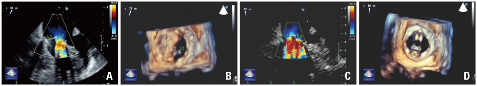
Fig. 1 Two dimensional transesophageal echocardiogram (2D TEE) showing one leaflet (L1) blocked, while the second leaflet (L2) is opening with accelerated color flow velocity (A). Real time three dimensional transesophageal echocardiogram (RT3DTEE) view from left atrium revealing one leaflet (L1) blocked, while the second leaflet (L2) is opening (B). 2D TEE after successful treatment leaflets excursion are normal with normal color flow velocity (C) and RT3DTEE (D).
Reference
-
1. Krim SR, Vivo RP, Patel A, Xu J, Igo SR, Zoghbi WA, Little SH. Direct assessment of normal mechanical mitral valve orifice area by real-time 3D echocardiography. JACC Cardiovasc Imaging. 2012; 5:478–483.
Article2. Lengyel M, Horstkotte D, Völler H, Mistiaen WP. Working Group Infection, Thrombosis, Embolism and Bleeding of the Society for Heart Valve Disease. Recommendations for the management of prosthetic valve thrombosis. J Heart Valve Dis. 2005; 14:567–575.
- Full Text Links
-
- Actions
-
Cited
- CITED
-
- Close
- Share
-
- Similar articles
-
- Echocardiography in Transcatheter Aortic Valve Implantation and Mitral Valve Clip
- Left Atrial Mural Endocarditis Diagnosed by Transesophageal Echocardiography in a Patient with Mitral Valve Prolapse
- Evaluation of functional regurgitation flow in patients with clinically normal mitral prosthesis by transesophageal echocardiography
- Tearing of the Mitral Valve during Vent Removal after a Successful Mitral Valve Repair: a Beneficial Role of Transesophageal Echocardiography
- Formation of intracardiac thrombus during cardiopulmonary bypass despite full heparinization and adequate activated clotting time: A case report
