Early Complication of Mustard Procedure after Late Repair
- Affiliations
-
- 1Department of Cardiovascular Diseases, Prince Salman Heart Center, King Fahad Medical City, Riyadh, Kingdom of Saudi Arabia. simustafa@kfmc.med.sa
- 2Department of Radiology, Prince Salman Heart Center, King Fahad Medical City, Riyadh, Kingdom of Saudi Arabia.
- 3Section of Pediatric Cardiology, University of Calgary, Calgary, AB, Canada.
- 4Adult Congenital Heart Disease Clinic, University of Calgary, Calgary, AB, Canada.
- 5Department of Pediatric Cardiology, Prince Salman Heart Center, King Fahad Medical City, Riyadh, Kingdom of Saudi Arabia.
- 6Division of Cardiovascular Diseases, Mayo Clinic Arizona, Scottsdale, AZ, USA.
- KMID: 2135422
- DOI: http://doi.org/10.4250/jcu.2013.21.4.200
Abstract
- No abstract available.
Keyword
MeSH Terms
Figure
-
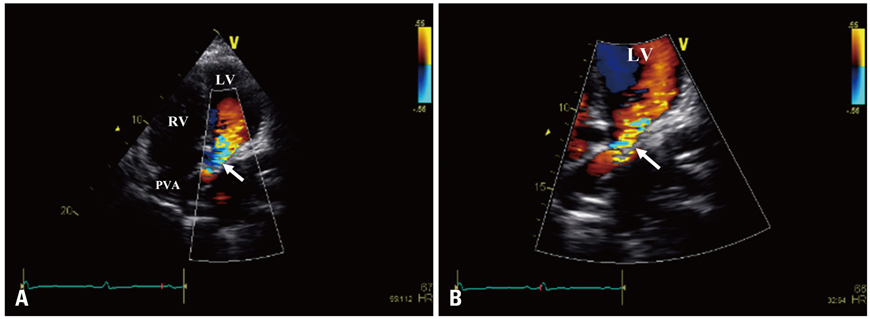
Fig. 1 Transthoracic echocardiogram apical 4-chamber view (A) with zoom mode (B) demonstrating the connections of the systemic venous circulation with significant color turbulence suggestive of baffle stenosis at the veno-atrial junction (arrow). LV: left ventricle, PVA: pulmonary venous atrium, RV: right ventricle.

Fig. 2 A: Cardiovascular magnetic resonance 4-chamber steady-state free precession (SSFP) image showing the trabeculated dilated systemic RV and patent pulmonary venous baffle (arrow). B: Cardiovascular magnetic resonance coronal SSFP image showing the typical discordant ventriculararterial relationship in complete transposition of the great arteries with the AO arising from the RV and MPA arising from the LV. C: Cardiovascular magnetic resonance axial SSFP image demonstrating severely dilated main PA and both branches. D: Cardiovascular magnetic resonance coronal SSFP image revealing significant stenosis of both superior vena cava (long arrow) and inferior vena cava (short arrow) limbs of the Mustard baffle at the veno-atrial junction. LV: left ventricle, PVA: pulmonary venous atrium, RV: right ventricle, AO: aorta, PA: pulmonary artery, LPA: left pulmonary artery, MPA: main pulmonary artery, RPA: right pulmonary artery.
Reference
-
1. Chatelain P, Meier B, Friedli B. Stenting of superior vena cava and inferior vena cava for symptomatic narrowing after repeated atrial surgery for D-transposition of the great vessels. Br Heart J. 1991; 66:466–468.
Article2. Cohen MD, Johnson T, Ramrakhiani S. MRI of surgical repair of transposition of the great vessels. AJR Am J Roentgenol. 2010; 194:250–260.
Article3. Kouchoukos NT, Blackstone EH, Doty DB, Hanley FL, Karp RB. Kirklin/Barratt-Boyes cardiac surgery. 3rd ed. Philadelphia: Churchill Livingstone;2003. p. 1439–1507.
- Full Text Links
-
- Actions
-
Cited
- CITED
-
- Close
- Share
-
- Similar articles
-
- Clinical Outcomes after Anatomic Repair Including Hemi-Mustard Operation in Patients with Congenitally Corrected Transposition of the Great Arteries
- Comparing of Complications of Inguinal Hernia Repair Using Prolene Hernia System
- Anatomical Repair of Congenitally Physiologically Corrected Transposition with Dextrocardia, Situs Inversus and the Interruption of Right Pulmonary Artery
- Outcome of Adult Hypospadias Repair
- Percutaneous Nephrolithotomy: Complication and Management
