Usefulness of Intraductal Ultrasonography in the Diagnosis of Cholangiocarcinoma and IgG4-Related Sclerosing Cholangitis
- Affiliations
-
- 1Department of Gastroenterology and Metabolism, Nagoya City University Graduate School of Medical Sciences, Nagoya, Japan. tnakazaw@med.nagoya-cu.ac.jp
Abstract
- The technique of intraductal ultrasonography (IDUS) of the bile duct with a thin-caliber probe and a ropeway system has provided excellent images of the bile duct and periductal structures and is an easy transpapillary approach. In addition, once the guide wire is inserted into the bile duct, IDUS and transpapillary biopsy after endoscopic retrograde cholangiopancreatography can be performed in a single session. Here, we review the usefulness of IDUS in the diagnosis of cholangiocarcinoma and IgG4-related sclerosing cholangitis.
MeSH Terms
Figure
-
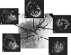
Fig. 1 Imagings of intraductal ultrasonography (IDUS) and cholangiogram of cholangiocarcinoma (infiltrating type). Cholagiogram showing stenosis in the hepatic hilum. (A) IDUS showing irrgular hypoechoic mass. (B) IDUS showing invasion to right hepatic artery. (C) IDUS showing irregular hypoechoic mass at the bifurcation of hepatic ducts. (D) No mass at the biurcation of B4. (E) IDUS showing heterogenous thickened wall in the middle of common bile duct.
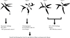
Fig. 2 Useful modalities in the diagnosis of IgG4-related sclerosing cholangitis based on shematic classification of cholangiogram. EUS-FNA, endoscopic ultrasonography-guided fine needle aspiration; UC, ulcerative colitis; IDUS, intraductal ultrasonography.
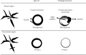
Fig. 3 Comparison of intraductal ultrasonography findings between IgG4-related sclerosing cholangitis and cholangiocarcinoma.
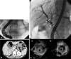
Fig. 4 Imagings of IgG4-related sclerosing cholangitis. (A) Endoscopic retrograde pancreatography with balloon catheter showing slight irregularities at the head of pancreas. (B) Abdominal computed tomography showing atrophic pancreas. (C) Endoscopic retrograde cholangiography (ERC) showing strictures in the both hepatic ducts at the hepatic hilum. Intraductal ultrasonography showing the homogeneous wall thickness with the size of 3.2 mm at the stenotic lesion of ERC (Ca) and with the size of 2.0 mm at the non-stenotic lesion of ERC (Cb).
Reference
-
1. Fujita N, Noda Y, Yokohata K, et al. Newly developed ultrasonic probe with ropeway for transpapillary intraductal ultrasonography of the bilio-pancreatic ductal system. Dig Endosc. 2000; 12:250–254.2. Menzel J, Domschke W. Gastrointestinal miniprobe sonography: the current status. Am J Gastroenterol. 2000; 95:605–616. PMID: 10710047.
Article3. Sakata J, Shirai Y, Wakai T, Nomura T, Sakata E, Hatakeyama K. Catheter tract implantation metastases associated with percutaneous biliary drainage for extrahepatic cholangiocarcinoma. World J Gastroenterol. 2005; 11:7024–7027. PMID: 16437610.
Article4. Tamada K, Nagai H, Yasuda Y, et al. Transpapillary intraductal US prior to biliary drainage in the assessment of longitudinal spread of extrahepatic bile duct carcinoma. Gastrointest Endosc. 2001; 53:300–307. PMID: 11231387.
Article5. Noda Y, Fujita N, Kobayashi G, et al. Intraductal ultrasonography before biliary drainage and transpapillary biopsy in assessment of the longitudinal extent of bile duct cancer. Dig Endosc. 2008; 20:73–78.
Article6. Nakazawa T, Ohara H, Sano H, Ando T, Joh T. Schematic classification of sclerosing cholangitis with autoimmune pancreatitis by cholangiography. Pancreas. 2006; 32:229. PMID: 16552350.
Article7. Nakazawa T, Ohara H, Sano H, et al. Difficulty in diagnosing autoimmune pancreatitis by imaging findings. Gastrointest Endosc. 2007; 65:99–108. PMID: 17185087.
Article8. Hayashi K, Nakazawa T, Ohara H, et al. Autoimmune sclerosing cholangiopancreatitis with little pancreatic involvements by imaging findings. Hepatogastroenterology. 2007; 54:2146–2151. PMID: 18251178.9. Nakazawa T, Naitoh I, Hayashi K, et al. Diagnostic criteria for IgG4-related sclerosing cholangitis based on cholangiographic classification. J Gastroenterol. 2012; 47:79–87. PMID: 21947649.
Article10. Naitoh I, Nakazawa T, Ohara H, et al. Endoscopic transpapillary intraductal ultrasonography and biopsy in the diagnosis of IgG4-related sclerosing cholangitis. J Gastroenterol. 2009; 44:1147–1155. PMID: 19636664.
Article11. Tamada K, Inui K, Menzel J. Intraductal ultrasonography of the bile duct system. Endoscopy. 2001; 33:878–885. PMID: 11605603.
Article12. Menzel J, Poremba C, Dietl KH, Domschke W. Preoperative diagnosis of bile duct strictures: comparison of intraductal ultrasonography with conventional endosonography. Scand J Gastroenterol. 2000; 35:77–82. PMID: 10672839.13. Tamada K, Ueno N, Tomiyama T, et al. Characterization of biliary strictures using intraductal ultrasonography: comparison with percutaneous cholangioscopic biopsy. Gastrointest Endosc. 1998; 47:341–349. PMID: 9609424.
Article14. Tamada K, Ido K, Ueno N, Kimura K, Ichiyama M, Tomiyama T. Preoperative staging of extrahepatic bile duct cancer with intraductal ultrasonography. Am J Gastroenterol. 1995; 90:239–246. PMID: 7847293.15. Yamaguchi K, Chijiiwa K, Saiki S, Shimizu S, Takashima M, Tanaka M. Carcinoma of the extrahepatic bile duct: mode of spread and its prognostic implications. Hepatogastroenterology. 1997; 44:1256–1261. PMID: 9356836.16. Sakamoto E, Nimura Y, Hayakawa N, et al. The pattern of infiltration at the proximal border of hilar bile duct carcinoma: a histologic analysis of 62 resected cases. Ann Surg. 1998; 227:405–411. PMID: 9527064.17. Nimura Y. Staging of biliary carcinoma: cholangiography and cholangioscopy. Endoscopy. 1993; 25:76–80. PMID: 8384103.
Article18. Nakazawa T, Ohara H, Sano H, et al. Cholangiography can discriminate sclerosing cholangitis with autoimmune pancreatitis from primary sclerosing cholangitis. Gastrointest Endosc. 2004; 60:937–944. PMID: 15605009.
Article19. Ghazale A, Chari ST, Zhang L, et al. Immunoglobulin G4-associated cholangitis: clinical profile and response to therapy. Gastroenterology. 2008; 134:706–715. PMID: 18222442.
Article
- Full Text Links
-
- Actions
-
Cited
- CITED
-
- Close
- Share
-
- Similar articles
-
- Immunoglobulin G4-related sclerosing cholangitis
- IgG4-Related Sclerosing Cholangitis and Primary Sclerosing Cholangitis
- Classification and Diagnostic Criteria for IgG4-Related Sclerosing Cholangitis
- Advances in IgG4-related Hepatobiliary Disease
- IgG4-related sclerosing cholangitis: liver biopsy findings
