Clin Endosc.
2012 Sep;45(3):324-327.
Usefulness of Endoscopic Ultrasound in Esophagogastric Varices
- Affiliations
-
- 1Department of Internal Medicine, Kyung Hee University School of Medicine, Seoul, Korea. joyshim@khu.ac.kr
Abstract
- Endoscopic ultrasound (EUS) is a useful diagnostic tool for evaluation of esophagogastric varices and guidance of endoscopic therapy. EUS can visualize not only collateral veins around the esophagus but also perforating veins that connect esophageal varices with collateral veins. They are associated with high risk of bleeding and early recurrence after initial variceal eradication. Isolated gastric varices can be easily diagnosed using EUS that mimic thickened gastric folds or subepithelial tumors. EUS-guided endoscopic therapy is a challenging field of variceal bleeding. It has a potential role for assistance of interventions and evaluation of treatment outcome.
MeSH Terms
Figure
-
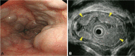
Fig. 1 Esophageal varices. (A) Endoscopic image shows large esophageal varices. (B) Endoscopic ultrasound image shows esophageal varices (arrowheads) using a miniature probe (20 MHz) in the same patient.
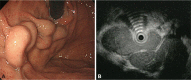
Fig. 2 Gastric varices. (A) Endoscopic image shows large varices in the stomach. (B) Endoscopic ultrasound image shows same gastric varices. A tortuous hypoechoic tubular structure is seen in the submucosal layer.

Fig. 3 Endoscopic ultrasound images of collateral and perforating veins. (A) A collateral vein outside of the esophageal wall is seen (arrow head). (B) A perforating vein (arrowhead) that connects a collateral vein with an esophageal varix is seen.
Reference
-
1. Garcia-Tsao G, Sanyal AJ, Grace ND, Carey W. Practice Guidelines Committee of the American Association for the Study of Liver Diseases. Practice Parameters Committee of the American College of Gastroenterology. Prevention and management of gastroesophageal varices and variceal hemorrhage in cirrhosis. Hepatology. 2007; 46:922–938. PMID: 17879356.
Article2. El-Saadany M, Jalil S, Irisawa A, Shibukawa G, Ohira H, Bhutani MS. EUS for portal hypertension: a comprehensive and critical appraisal of clinical and experimental indications. Endoscopy. 2008; 40:690–696. PMID: 18609464.
Article3. Irisawa A, Saito A, Obara K, et al. Endoscopic recurrence of esophageal varices is associated with the specific EUS abnormalities: severe periesophageal collateral veins and large perforating veins. Gastrointest Endosc. 2001; 53:77–84. PMID: 11154493.
Article4. Sato T, Yamazaki K, Toyota J, et al. Perforating veins in recurrent esophageal varices evaluated by endoscopic color Doppler ultrasonography with a galactose-based contrast agent. J Gastroenterol. 2004; 39:422–428. PMID: 15175939.
Article5. Miller LS, Schiano TD, Adrain A, et al. Comparison of high-resolution endoluminal sonography to video endoscopy in the detection and evaluation of esophageal varices. Hepatology. 1996; 24:552–555. PMID: 8781323.
Article6. Miller L, Banson FL, Bazir K, et al. Risk of esophageal variceal bleeding based on endoscopic ultrasound evaluation of the sum of esophageal variceal cross-sectional surface area. Am J Gastroenterol. 2003; 98:454–459. PMID: 12591068.
Article7. Schiano TD, Adrain AL, Vega KJ, Liu JB, Black M, Miller LS. High-resolution endoluminal sonography assessment of the hematocystic spots of esophageal varices. Gastrointest Endosc. 1999; 49(4 Pt 1):424–427. PMID: 10202053.
Article8. Wu CS, Chen CM, Chang KY. Endoscopic injection sclerotherapy of bleeding duodenal varices. J Gastroenterol Hepatol. 1995; 10:481–483. PMID: 8527718.
Article9. Faigel DO, Rosen HR, Sasaki A, Flora K, Benner K. EUS in cirrhotic patients with and without prior variceal hemorrhage in comparison with noncirrhotic control subjects. Gastrointest Endosc. 2000; 52:455–462. PMID: 11023560.
Article10. Sato T, Yamazaki K, Toyota J, Karino Y, Ohmura T, Akaike J. Endoscopic ultrasonographic evaluation of hemodynamics related to variceal relapse in esophageal variceal patients. Hepatol Res. 2009; 39:126–133. PMID: 19208033.
Article11. Hino S, Kakutani H, Ikeda K, et al. Hemodynamic assessment of the left gastric vein in patients with esophageal varices with color Doppler EUS: factors affecting development of esophageal varices. Gastrointest Endosc. 2002; 55:512–517. PMID: 11923763.
Article12. Salama ZA, Kassem AM, Giovannini M, Hunter MS. Endoscopic ultrasonographic study of the azygos vein in patients with varices. Endoscopy. 1997; 29:748–750. PMID: 9427495.
Article13. Kassem AM, Salama ZA, Zakaria MS, Hassaballah M, Hunter MS. Endoscopic ultrasonographic study of the azygos vein before and after endoscopic obliteration of esophagogastric varices by injection sclerotherapy. Endoscopy. 2000; 32:630–634. PMID: 10935792.
Article14. Irisawa A, Obara K, Sato Y, et al. EUS analysis of collateral veins inside and outside the esophageal wall in portal hypertension. Gastrointest Endosc. 1999; 50:374–380. PMID: 10462659.
Article15. Obara K. Hemodynamic mechanism of esophageal varices. Dig Endosc. 2006; 18:6–9.
Article16. Nakamura S, Murata Y, Mitsunaga A, Oi I, Hayashi N, Suzuki S. Hemodynamics of esophageal varices on three-dimensional endoscopic ultrasonography and indication of endoscopic variceal ligation. Dig Endosc. 2003; 15:289–297.
Article17. Leung VK, Sung JJ, Ahuja AT, et al. Large paraesophageal varices on endosonography predict recurrence of esophageal varices and rebleeding. Gastroenterology. 1997; 112:1811–1816. PMID: 9178670.
Article18. Lo GH, Lai KH, Cheng JS, Huang RL, Wang SJ, Chiang HT. Prevalence of paraesophageal varices and gastric varices in patients achieving variceal obliteration by banding ligation and by injection sclerotherapy. Gastrointest Endosc. 1999; 49(4 Pt 1):428–436. PMID: 10202054.
Article19. Suzuki T, Matsutani S, Umebara K, et al. EUS changes predictive for recurrence of esophageal varices in patients treated by combined endoscopic ligation and sclerotherapy. Gastrointest Endosc. 2000; 52:611–617. PMID: 11060184.
Article20. Hino S, Kakutani H, Ikeda K, et al. Hemodynamic analysis of esophageal varices using color Doppler endoscopic ultrasonography to predict recurrence after endoscopic treatment. Endoscopy. 2001; 33:869–872. PMID: 11571684.
Article21. Lahoti S, Catalano MF, Alcocer E, Hogan WJ, Geenen JE. Obliteration of esophageal varices using EUS-guided sclerotherapy with color Doppler. Gastrointest Endosc. 2000; 51:331–333. PMID: 10699783.
Article22. de Paulo GA, Ardengh JC, Nakao FS, Ferrari AP. Treatment of esophageal varices: a randomized controlled trial comparing endoscopic sclerotherapy and EUS-guided sclerotherapy of esophageal collateral veins. Gastrointest Endosc. 2006; 63:396–402. PMID: 16500386.23. Binmoeller KF, Weilert F, Shah JN, Kim J. EUS-guided transesophageal treatment of gastric fundal varices with combined coiling and cyanoacrylate glue injection (with videos). Gastrointest Endosc. 2011; 74:1019–1025. PMID: 21889139.
Article
- Full Text Links
-
- Actions
-
Cited
- CITED
-
- Close
- Share
-
- Similar articles
-
- Post-TIPS Change of Esophagogastric Variceal Size on Endoscopy
- Endoscopic Observation of Gastric Varices
- A case of bleeding duodenal varix treated by endoscopic band ligation
- Practical Approach to Endoscopic Management for Bleeding Gastric Varices
- Endoscopic Treatment and Prevention of Acute Variceal Hemorrhage
