J Korean Assoc Pediatr Surg.
2015 Dec;21(2):42-45. 10.13029/jkaps.2015.21.2.42.
Mature Cystic Gastric Teratoma in an Infant: A Case Presenting with a Gastrointestinal Bleeding
- Affiliations
-
- 1Department of Pediatric Surgery, Pusan National University Yangsan Hospital, Yangsan, Korea. choyh70@pusan.ac.kr
- 2Department of Pediatrics, Pusan National University Yangsan Hospital, Yangsan, Korea.
- KMID: 2133790
- DOI: http://doi.org/10.13029/jkaps.2015.21.2.42
Abstract
- Gastric teratoma is an extremely rare tumor that accounts for less than 1% of all teratomas. Gastric teratoma is mostly presented as a palpable abdominal mass, and is rarely accompanied with gastrointestinal bleeding such as melena or hematemesis. A 5-month-old male infant was brought with a history of pale facial color and dark-colored stool. The hemoglobin level was at 6.1 g/dL, with melena having begun 1 month previous. Upper gastrointestinal endoscopy revealed a polypoid mass with bleeding at the upper body and lesser curvature of the stomach. Wedge resection of the stomach was performed and histopathological analysis confirmed the mass to be a mature cystic teratoma. There was no recurrence after the operation during follow-up.
Keyword
MeSH Terms
Figure
-
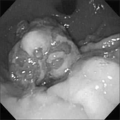
Fig. 1 Endoscopic finding shows multi-lobulated polypoid mass with oozing at upper body of stomach.

Fig. 2 Gross finding shows 4.2×3.4×3.0 cm sized multi-lobulated mass, with both exo- and endophytic growth.
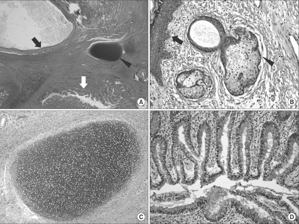
Fig. 3 Microscopic findings. (A) All 3 germ layer tissues; black arrow, squamous epithelium lined cystic lesion from ectoderm; black arrowhead, cartilage from mesoderm; white arrow, small intestinal epithelium from endoderm (H&E, ×40). (B) Ectoderm origin tissues; black arrow, squamous epithelium; black arrowhead, sebaceous gland (H&E, ×100). (C) Mesoderm origin tissue; cartilage (H&E, ×100). (D) Endoderm origin tissue; gastrointestinal epithelium (H&E, ×100).
Reference
-
1. Wakhlu A, Wakhlu AK. Paediatric gastric teratoma. Eur J Pediatr Surg. 2002; 12:375–378.2. Anilkumar MG, Jagadishkumar K, Girish GN, Sunila . Immature gastric teratoma in an infant. Indian J Surg. 2013; 75:Suppl 1. 453–455.3. Yoon SE, Goo HW, Jun S, Lee IC, Yoon CH. Immature gastric teratoma in an infant: a case report. Korean J Radiol. 2000; 1:226–228.4. Yaji PR, Joshi S, Kinhal V, Ravishankar TH, Jayaprakasha G, Melapure A, et al. Gastric teratoma in an infant: a rare case report and discussion. Indian J Surg. 2013; 75:Suppl 1. 152–154.5. Eusterman GB, Sentry EG. Benign tumours of the stomach: report of 27 cases. Surg Gynecol Obstet. 1922; 34:372–378.6. Gupta V, Babu RY, Rana S, Vaiphei K, Rao KL, Bhasin DK. Mature gastric teratoma: recurrence in adulthood. J Pediatr Surg. 2009; 44:e17–e19.7. Saha M. Malignant gastric teratoma: report of two cases from a single center. Pediatr Surg Int. 2010; 26:931–934.8. Kumar V, Godara R, Bharadwaj R, Arora M. Gastric teratoma-unusual cause of neonatal obstructive jaundice: a case report. Indian J Surg. 2013; 75:Suppl 1. 421–424.9. Jeong HC, Cha SJ, Kim GJ. Rapidly grown congenital fetal immature gastric teratoma causing severe neonatal respiratory distress. J Obstet Gynaecol Res. 2012; 38:449–451.10. Gupta DK, Srinivas M, Dave S, Agarwala S, Bajpai M, Mitra DK. Gastric teratoma in children. Pediatr Surg Int. 2000; 16:329–332.
- Full Text Links
-
- Actions
-
Cited
- CITED
-
- Close
- Share
-
- Similar articles
-
- Exogastric Mature Teratoma in an Infant: A Case Report
- A Case of Squamous Cell Carcinoma Arising in the Mature Cystic Teratoma with Direct Invasion to Transverse Colon and Jejunum
- Traumatic Rupture of a Mature Cystic Teratoma of the Ovary
- A Case of Papillary Carcinoma of Thyroid Gland Arising from Ovarian Mature Cystic Teratoma
- A Case of Delayed Operated Huge Mature Cystic Teratoma of the Ovary
