Radiolucent rim as a possible diagnostic aid for differentiating jaw lesions
- Affiliations
-
- 1Department of Oral Medicine, School of Dentistry, Shahid Beheshti University of Medical Sciences, Tehran, Iran. m-baharvand@sbmu.ac.ir
- KMID: 2132939
- DOI: http://doi.org/10.5624/isd.2015.45.4.253
Abstract
- In this study, we formulate a new proposal that complements previous classifications in order to assist dental practitioners in performing a differential diagnosis based on patients' radiographs. We used general search engines and specialized databases such as Google Scholar, PubMed, PubMed Central, MedLine Plus, Science Direct, Scopus, and well-recognized textbooks to find relevant studies by using keywords such as "jaw disease," "jaw lesions," "radiolucent rim," "radiolucent border," and "radiolucent halo." More than 200 articles were found, of which 70 were broadly relevant to the topic. We ultimately included 50 articles that were closely related to the topic of interest. When the relevant data were compiled, the following eight lesions were identified as having a radiolucent rim: periapical cemento-osseous dysplasia, focal cemento-osseous dysplasia, florid cemento-osseous dysplasia, cemento-ossifying fibroma, osteoid osteoma, osteoblastoma, odontoma, and cementoblastoma. We propose a novel subcategory, jaw lesions with a radiolucent rim, which includes eight entities. The implementation of this new category can help improve the diagnoses that dental practitioners make based on patients' radiographs.
Keyword
MeSH Terms
Figure
-
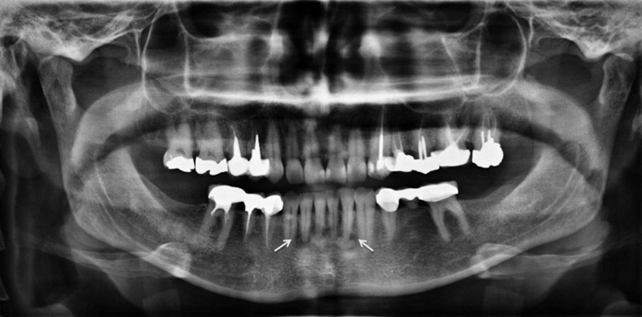
Fig. 1 A panoramic radiograph shows a radiolucent rim at the periphery of lesions (arrows) diagnosed as periapical cemento-osseous dysplasia.

Fig. 2 Focal cemento-osseous dysplasia is seen at the periapex of the left mandibular first molar with a radiolucent rim (arrow) on a panoramic radiograph.

Fig. 3 Florid cemento-osseous dysplasia is situated at the right, left, and anterior portions of the mandible, surrounded by a radiolucent rim (arrows).
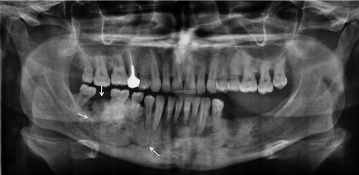
Fig. 4 A very large cemento-ossifying fibroma is located in the right aspect of the mandible with a radiolucent rim around the lesion (arrow).

Fig. 5 A complex odontoma with a radiolucent rim in conjunction with a corticated border around the lesion (arrows).

Fig. 6 Cementoblastoma is seen at the periapex of the left second mandibular molar. A radiolucent halo is apparent at the periphery of the lesion (arrows).
Cited by 4 articles
-
Jaw lesions associated with impacted tooth: A radiographic diagnostic guide
Hamed Mortazavi, Maryam Baharvand
Imaging Sci Dent. 2016;46(3):147-157. doi: 10.5624/isd.2016.46.3.147.Review of common conditions associated with periodontal ligament widening
Hamed Mortazavi, Maryam Baharvand
Imaging Sci Dent. 2016;46(4):229-237. doi: 10.5624/isd.2016.46.4.229.Recurrent symptomatic cemento-osseous dysplasia: A case report
Chang-Ki Min, Kwang-Joon Koh, Kyoung-A Kim
Imaging Sci Dent. 2018;48(2):131-137. doi: 10.5624/isd.2018.48.2.131.Common conditions associated with displacement of the inferior alveolar nerve canal: A radiographic diagnostic aid
Hamed Mortazavi, Maryam Baharvand, Yaser Safi, Mohammad Behnaz
Imaging Sci Dent. 2019;49(2):79-86. doi: 10.5624/isd.2019.49.2.79.
Reference
-
1. Neyaz Z, Gadodia A, Gamanagatti S, Mukhopadhyay S. Radiographical approach to jaw lesions. Singapore Med J. 2008; 49:165–177.2. George G, Padiyath S. Unicystic jaw lesions: a radiographic guideline. J Indian Acad Oral Med Radiol. 2010; 22:S31–S36.
Article3. Curé JK, Vattoth S, Shah R. Radiopaque jaw lesions: an approach to the differential diagnosis. Radiographics. 2012; 32:1909–1925.
Article4. Wood NK, Goaz PW. Differential diagnosis of oral and maxillofacial lesions. 5th ed. St Louis: Mosby-Year Book Inc.;1997. p. 415–448.5. Sanjai K, Kumarswamy J, Kumar VK, Patil A. Florid cemento osseous dysplasia in association with dentigerous cyst. J Oral Maxillofac Pathol. 2010; 14:63–68.
Article6. Barnes L, Eveson JW, Reichart P, Sidransky D. Pathology and genetics of head and neck tumours. World Health Organization Classification of Tumours. Lyon: IARC Press;2005.7. Alsufyani NA, Lam EW. Osseous (cement-osseous) dysplasia of the jaws: clinical and radiographic analysis. J Can Dent Assoc. 2011; 77:b70.8. Kawai T, Hiranuma H, Kishino M, Jikko A, Sakuda M. Cemento-osseous dysplasia of the jaws in 54 Japanese patients: a radiographic study. Oral Surg Oral Med Oral Pathol Oral Radiol Endod. 1999; 87:107–114.9. Alsufyani NA, Lam EW. Cemento-osseous dysplasia of the jaw bones: key radiographic features. Dentomaxillofac Radiol. 2011; 40:141–146.
Article10. Beylouni I, Farge P, Mazoyer JF, Coudert JL. Florid cemento-osseous dysplasia: report of a case documented with computed tomography and 3D imaging. Oral Surg Oral Med Oral Pathol Oral Radiol Endod. 1998; 85:707–711.11. Brannon RB, Fowler CB. Benign fibro-osseous lesions: a review of current concepts. Adv Anat Pathol. 2001; 8:126–143.
Article12. Eskandarloo A, Yousefi F. CBCT findings of periapical cemento-osseous dysplasia: a case report. Imaging Sci Dent. 2013; 43:215–218.
Article13. Zegarelli E, Kutscher A, Napoli N, Iurono F, Hoffman P. The cementoma. A study of 230 patients with 435 cementomas. Oral Surg Oral Med Oral Pathol. 1964; 17:219–224.14. Komabayashi T, Zhu Q. Cemento-osseous dysplasia in an elderly Asian male: a case report. J Oral Sci. 2011; 53:117–120.
Article15. Summerlin DJ, Tomich CE. Focal cemento-osseous dysplasia: a clinicopathologic study of 221 cases. Oral Surg Oral Med Oral Pathol. 1994; 78:611–620.
Article16. Bhandari R, Sandhu SV, Bansal H, Behl R, Bhullar RK. Focal cemento-osseous dysplasia masquerading as a residual cyst. Contemp Clin Dent. 2012; 3:Suppl 1. S60–S62.
Article17. Eversole R, Su L, ElMofty S. Benign fibro-osseous lesions of the craniofacial complex. A review. Head Neck Pathol. 2008; 2:177–202.
Article18. Rao GS, Kamalapur MG, Acharya S. Focal cemento-osseous dysplasia masquerading as benign cementoblastoma: a diagnostic dilemma. J Oral Maxillofac Pathol. 2014; 18:150.
Article19. Köse TE, Köse OD, Karabas HC, Erdem TL. Ozcan I. Findings of florid cemento-osseous dysplasia: a report of three cases. J Oral Maxillofac Res. 2014; 4:e4.20. Kutluay Köklü H, Cankal DA, Bozkaya S, Ergün G, Bar E. Florid cemento-osseous dysplasia: report of a case documented with clinical, radiographic, biochemical and histological findings. J Clin Exp Dent. 2013; 5:e58–e61.
Article21. K L K, R S AB, P S, Kumaran S. Pericoronal occurrence of cemento-ossifying fibroma: an unexemplified and unusual case report with review of literature. J Clin Diagn Res. 2014; 8:277–279.22. Chang CC, Hung HY, Chang JY, Yu CH, Wang YP, Liu BY, et al. Central ossifying fibroma: a clinicopathologic study of 28 cases. J Formos Med Assoc. 2008; 107:288–294.
Article23. Trijolet JP, Parmentier J, Sury F, Goga D, Mejean N, Laure B. Cemento-ossifying fibroma of the mandible. Eur Ann Otorhinolaryngol Head Neck Dis. 2011; 128:30–33.
Article24. Khan SA, Sharma NK, Raj V, Sethi T. Ossifying fibroma of maxilla in a male child: report of a case and review of the literature. Natl J Maxillofac Surg. 2011; 2:73–79.
Article25. Ida M, Kurabayashi T, Takahashi Y, Takagi M, Sasaki T. Osteoid osteoma in the mandible. Dentomaxillofac Radiol. 2002; 31:385–387.
Article26. An SY, Shin HI, Choi KS, Park JW, Kim YG, Benavides E, et al. Unusual osteoid osteoma of the mandible: report of case and review of the literature. Oral Surg Oral Med Oral Pathol Oral Radiol. 2013; 116:e134–e140.
Article27. Liu CJ, Chang KW, Chang KM, Cheng CY. A variant of osteoid osteoma of the mandible: report of a case. J Oral Maxillofac Surg. 2002; 60:219–221.
Article28. Manjunatha BS, Sunit P, Amit M, Sanjiv S. Osteoblastoma of the jaws: report of a case and review of literature. Clin Pract. 2011; 1:e118.
Article29. Alvares Capelozza AL, Gião Dezotti MS, Casati Alvares L, Negrão Fleury R, Sant'Ana E. Osteoblastoma of the mandible: systematic review of the literature and report of a case. Dentomaxillofac Radiol. 2005; 34:1–8.
Article30. Bokhari K, Hameed MS, Ajmal M, Togoo RA. Benign osteoblastoma involving maxilla: a case report and review of the literature. Case Rep Dent. 2012; 2012:351241.
Article31. Jones AC, Prihoda TJ, Kacher JE, Odingo NA, Freedman PD. Osteoblastoma of the maxilla and mandible: a report of 24 cases, review of the literature, and discussion of its relationship to osteoid osteoma of the jaws. Oral Surg Oral Med Oral Pathol Oral Radiol Endod. 2006; 102:639–650.
Article32. Oztürk M, Ozeç I, Aker H, Müslehiddinoğlu A. Osteoblastoma of the mandible with root resorption: a case report. Quintessence Int. 2003; 34:135–138.33. Shah S, Kim JE, Huh KH, Yi WJ, Heo MS, Lee SS. Recurrent osteoblastoma of the maxilla. Dentomaxillofac Radiol. 2013; 42:20100263.
Article34. Junquera L, de Vicente JC, Roig P, Olay S, Rodríguez-Recio O. Intraosseous odontoma erupted into the oral cavity: an unusual pathology. Med Oral Patol Oral Cir Bucal. 2005; 10:248–251.35. Piattelli A, Perfetti G, Carraro A. Complex odontoma as a periapical and interradicular radiopacity in a primary molar. J Endod. 1996; 22:561–563.
Article36. Reddy GS, Reddy GV, Sidhartha B, Sriharsha K, Koshy J, Sultana R. Large complex odontoma of mandible in a young boy: a rare and unusual case report. Case Rep Dent. 2014; 2014:854986.
Article37. Ohtawa Y, Ichinohe S, Kimura E, Hashimoto S. Erupted complex odontoma delayed eruption of permanent molar. Bull Tokyo Dent Coll. 2013; 54:251–257.
Article38. Hidalgo-Sánchez O, Leco-Berrocal MI, Martínez-González JM. Metaanalysis of the epidemiology and clinical manifestations of odontomas. Med Oral Patol Oral Cir Bucal. 2008; 13:E730–E734.39. Philipsen HP, Reichart PA, Praetorius F. Mixed odontogenic tumours and odontomas. Considerations on interrelationship. Review of the literature and presentation of 134 new cases of odontomas. Oral Oncol. 1997; 33:86–99.
Article40. Garcia-Consuegra L, Junquera LM, Albertos JM, Rodriguez . Odontomas. A clinical-histological and retrospective epidemiological study of 46 cases. Med Oral. 2000; 5:367–372.41. Sharma N. Benign cementoblastoma: a rare case report with review of literature. Contemp Clin Dent. 2014; 5:92–94.
Article42. Sankari LS, Ramakrishnan K. Benign cementoblastoma. J Oral Maxillofac Pathol. 2011; 15:358–360.
Article43. Kumar S, Prabhakar V, Angra R. Infected cementoblastoma. Natl J Maxillofac Surg. 2011; 2:200–203.
Article44. Gulses A, Bayar GR, Aydin C, Sencimen M. A case of a benign cementoblastoma treated by enucleation and apicoectomy. Gen Dent. 2012; 60:e380–e382.45. Iannaci G, Luise R, Iezzi G, Piattelli A, Salierno A. Multiple cementoblastoma: a rare case report. Case Rep Dent. 2013; 2013:828373.
Article46. Brannon RB, Fowler CB, Carpenter WM, Corio RL. Cementoblastoma: an innocuous neoplasm? A clinicopathologic study of 44 cases and review of the literature with special emphasis on recurrence. Oral Surg Oral Med Oral Pathol Oral Radiol Endod. 2002; 93:311–320.
Article47. Huber AR, Folk GS. Cementoblastoma. Head Neck Pathol. 2009; 3:133–135.
Article48. Bilodeau E, Collins B, Costello B, Potluri A. Case report: a pediatric case of cementoblastoma with histologic and radiographic features of an osteoblastoma and osteosarcoma. Head Neck Pathol. 2010; 4:324–328.
Article49. Harada H, Omura K, Mogi S, Okada N. Cementoblastoma arising in the maxilla of an 8-year-old boy: a case report. Int J Dent. 2011; 2011:384578.
Article50. Pacifici L, Tallarico M, Bartoli A, Ripari A, Cicconetti A. Benign cementoblastoma: a clinical case of conservative surgical treatment of the involved tooth. Minerva Stomatol. 2004; 53:685–691.51. Monks FT, Bradley JC, Turner EP. Central osteoblastoma or cementoblastoma? A case report and 12 year review. Br J Oral Surg. 1981; 19:29–37.
Article
- Full Text Links
-
- Actions
-
Cited
- CITED
-
- Close
- Share
-
- Similar articles
-
- Scalloped border as a possible diagnostic aid for differentiating jaw lesions: A pictorial essay
- Differential diagnosis between odontogenic keratocyst and ameloblastoma by computed tomography
- Pinhole Bone Scintigraphic Manifestation of Fibrous Dysplasia
- A clinical study on fibro-osseous lesions of the jaws
- A study on the mixed jaw lesions associated with teeth
