Imaging Sci Dent.
2015 Dec;45(4):213-220. 10.5624/isd.2015.45.4.213.
Cephalometric landmark variability among orthodontists and dentomaxillofacial radiologists: a comparative study
- Affiliations
-
- 1Department of Dental Radiology, Faculty of Dental Medicine, University of Porto, Porto, Portugal. paula.o.reis@gmail.com
- 2Department of Surgery, Dentistry School, Pontifical Catholic University of Rio Grande do Sul, Porto Alegre, Rio Grande do Sul, Brazil.
- 3Department of Radiology, Faculty of Dentistry, Chulalongkorn University, Bangkok, Thailand.
- 4Department of Clinical Dentistry, Faculty of Health Science, UiT The Arctic University of Norway, Tromso, Norway.
- 5Department of Orthodontics, Faculty of Dental Medicine, University of Porto, Portugal.
- 6Oral Imaging Center, OMFS-IMPATH Research Group, Department of Imaging and Pathology, Faculty of Medicine, University of Leuven, Leuven, Belgium.
- KMID: 2132933
- DOI: http://doi.org/10.5624/isd.2015.45.4.213
Abstract
- PURPOSE
The aim this study was to compare the accuracy of orthodontists and dentomaxillofacial radiologists in identifying 17 commonly used cephalometric landmarks, and to determine the extent of variability associated with each of those landmarks.
MATERIALS AND METHODS
Twenty digital lateral cephalometric radiographs were evaluated by two groups of dental specialists, and 17 cephalometric landmarks were identified. The x and y coordinates of each landmark were recorded. The mean value for each landmark was considered the best estimate and used as the standard. Variation in measurements of the distance between landmarks and measurements of the angles associated with certain landmarks was also assessed by a subset of two observers, and intraobserver and interobserver agreement were evaluated.
RESULTS
Intraclass correlation coefficients were excellent for intraobserver agreement, but only good for interobserver agreement. The least reliable landmark for orthodontists was the gnathion (Gn) point (standard deviation [SD], 5.92 mm), while the orbitale (Or) was the least reliable landmark (SD, 4.41 mm) for dentomaxillofacial radiologists. Furthermore, the condylion (Co)-Gn plane was the least consistent (SD, 4.43 mm).
CONCLUSION
We established that some landmarks were not as reproducible as others, both horizontally and vertically. The most consistently identified landmark in both groups was the lower incisor border, while the least reliable points were Co, Gn, Or, and the anterior nasal spine. Overall, a lower level of reproducibility in the identification of cephalometric landmarks was observed among orthodontists.
MeSH Terms
Figure
-
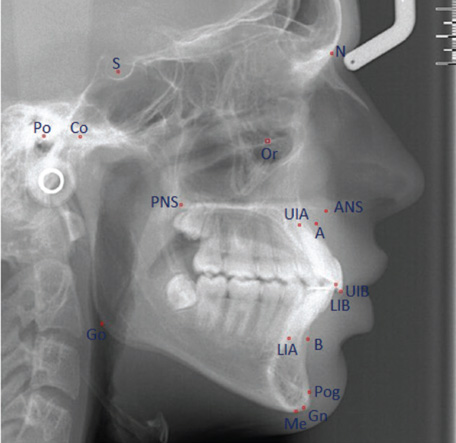
Fig. 1 Cephalometric landmarks used in the study. N, nasion; Or, orbitale; S, sella; Co, condylion; Po, porion, PNS, posterior nasal spine; ANS, anterior nasal spine; A, point A; UIA, upper incisor apex; UIB, upper incisor border; LIB, lower incisor border; LIA, lower incisor apex; B, point B; Pog, pogonion; Gn, gnathion; Me, menton; Go, gonion.

Fig. 2 Example of a lateral cephalometric radiograph with the identification of landmarks by two observers.
Reference
-
1. Broadbent BH. A new x-ray technique and its application to orthodontia. Angle Orthod. 1931; 1:45–66.2. Baumrind S, Frantz RC. The reliability of head film measurements. 1. Landmark identification. Am J Orthod. 1971; 60:111–127.3. Chen YJ, Chen SK, Huang HW, Yao CC, Chang HF. Reliability of landmark identification in cephalometric radiography acquired by a storage phosphor imaging system. Dentomaxillofac Radiol. 2004; 33:301–306.
Article4. Kamoen A, Dermaut L, Verbeeck R. The clinical significance of error measurement in the interpretation of treatment results. Eur J Orthod. 2001; 23:569–578.
Article5. Miloro M, Borba AM, Ribeiro-Junior O, Naclério-Homem MG, Jungner M. Is there consistency in cephalometric landmark identification amongst oral and maxillofacial surgeons? Int J Oral Maxillofac Surg. 2014; 43:445–453.
Article6. Tng TT, Chan TC, Hägg U, Cooke MS. Validity of cephalometric landmarks. An experimental study on human skulls. Eur J Orthod. 1994; 16:110–120.
Article7. Gravely JF, Benzies PM. The clinical significance of tracing error in cephalometry. Br J Orthod. 1974; 1:95–101.
Article8. Kvam E, Krogstad O. Variability in tracings of lateral head plates for diagnostic orthodontic purposes. A methodologic study. Acta Odontol Scand. 1969; 27:359–369.9. Lau PY, Cooke MS, Hägg U. Effect of training and experience on cephalometric measurement errors on surgical patients. Int J Adult Orthodon Orthognath Surg. 1997; 12:204–213.10. da Silveira HL, Silveira HE. Reproducibility of cephalometric measurements made by three radiology clinics. Angle Orthod. 2006; 76:394–399.11. Proffit WR, Fields HW, Sarver DM. Contemporary orthodontics. 4th ed. St. Louis: Mosby Elsevier;2006.12. Shrout PE, Fleiss JL. Intraclass correlations: uses in assessing rater reliability. Psychol Bull. 1979; 86:420–428.
Article13. Ricketts RM. Analysis - the Interim. Angle Orthod. 1970; 40:129–137.14. McNamara JA Jr. A method of cephalometric evaluation. Am J Orthod. 1984; 86:449–469.
Article15. McClure SR, Sadowsky PL, Ferreira A, Jacobson A. Reliability of digital versus conventional cephalometric radiology: a comparative evaluation of landmark identification error. Semin Orthod. 2005; 11:98–110.
Article16. Shaheed S, Iftikhar A, Rasool G, Bashir U. Accuracy of linear cephalometric measurements with scanned lateral cephalograms. Pak Oral Dental J. 2011; 31:68–72.17. Murali RV, Sukumar MR, Tajir TF, Rajalingam S. Comparative study of manual cephalometric tracing and computerized cephalometric tracing in digital lateral cephalogram for accuracy and reliability of landmarks. Indian J Multidiscip Dent. 2011; 1:126–134.18. Durão AR, Pittayapat P, Rockenbach MI, Olszewski R, Ng S, Ferreira AP, et al. Validity of 2D lateral cephalometry in orthodontics: a systematic review. Prog Orthod. 2013; 14:31.
Article19. Houston WJ, Maher RE, McElroy D, Sherriff M. Sources of error in measurements from cephalometric radiographs. Eur J Orthod. 1986; 8:149–151.
Article20. Chien PC, Parks ET, Eraso F, Hartsfield JK, Roberts WE, Ofner S. Comparison of reliability in anatomical landmark identification using two-dimensional digital cephalometrics and three-dimensional cone beam computed tomography in vivo. Dentomaxillofac Radiol. 2009; 38:262–273.21. Medelnik J, Hertrich K, Steinhäuser-Andresen S, Hirschfelder U, Hofmann E. Accuracy of anatomical landmark identification using different CBCT- and MSCT-based 3D images: an in vitro study. J Orofac Orthop. 2011; 72:261–278.22. Lagravère MO, Low C, Flores-Mir C, Chung R, Carey JP, Heo G, et al. Intraexaminer and interexaminer reliabilities of landmark identification on digitized lateral cephalograms and formatted 3-dimensional cone-beam computerized tomography images. Am J Orthod Dentofacial Orthop. 2010; 137:598–604.
Article
- Full Text Links
-
- Actions
-
Cited
- CITED
-
- Close
- Share
-
- Similar articles
-
- The comparison of landmark identification errors and reproducibility between conventional lateral cephalometric radiography and digital lateral cephalometric radiography
- Reproducibility of Lateral Cephalometric Landmarks According to Radiographic Image Enhancement
- A cephalometric analysis on esthetic facial soft tissue of Korean young adult female
- Accuracy of Automatic Cephalometric Analysis Programs on Lateral Cephalograms of Preadolescent Children
- A comparative study of computed radiographic cephalometry and conventional cephalometry in reliability of head film measurements (landmarks identification)
