Imaging Sci Dent.
2015 Dec;45(4):205-211. 10.5624/isd.2015.45.4.205.
Comparison of micro-computerized tomography and cone-beam computerized tomography in the detection of accessory canals in primary molars
- Affiliations
-
- 1Department of Dentomaxillofacial Radiology, Faculty of Dentistry, Ankara University, Ankara, Turkey. dtbuketonder@gmail.com
- 2Department of Periodontology, Faculty of Dentistry, Hacettepe University, Ankara, Turkey.
- 3Department of Anatomy, Faculty of Medicine, Hacettepe University, Ankara, Turkey.
- 4Department of Pediatric Dentistry, Faculty of Dentistry, Kirikkale University, Kirikkale, Turkey.
- 5Department of Biostatistics, Faculty of Medicine, Yildirim Beyazit University, Ankara, Turkey.
- 6Department of Dentomaxillofacial Radiology, Gulhane Military Hospital, Dental Clinics, Ankara, Turkey.
- KMID: 2132932
- DOI: http://doi.org/10.5624/isd.2015.45.4.205
Abstract
- PURPOSE
This study was performed to compare the accuracy of micro-computed tomography (CT) and cone-beam computed tomography (CBCT) in detecting accessory canals in primary molars.
MATERIALS AND METHODS
Forty-one extracted human primary first and second molars were embedded in wax blocks and scanned using micro-CT and CBCT. After the images were taken, the samples were processed using a clearing technique and examined under a stereomicroscope in order to establish the gold standard for this study. The specimens were classified into three groups: maxillary molars, mandibular molars with three canals, and mandibular molars with four canals. Differences between the gold standard and the observations made using the imaging methods were calculated using Spearman's rho correlation coefficient test.
RESULTS
The presence of accessory canals in micro-CT images of maxillary and mandibular root canals showed a statistically significant correlation with the stereomicroscopic images used as a gold standard. No statistically significant correlation was found between the CBCT findings and the stereomicroscopic images.
CONCLUSION
Although micro-CT is not suitable for clinical use, it provides more detailed information about minor anatomical structures. However, CBCT is convenient for clinical use but may not be capable of adequately analyzing the internal anatomy of primary teeth.
Keyword
MeSH Terms
Figure
-
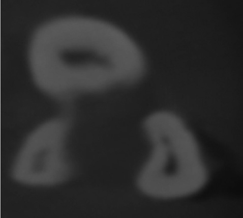
Fig. 1 The accessory canals are seen on an axial cone-beam computed tomography image.
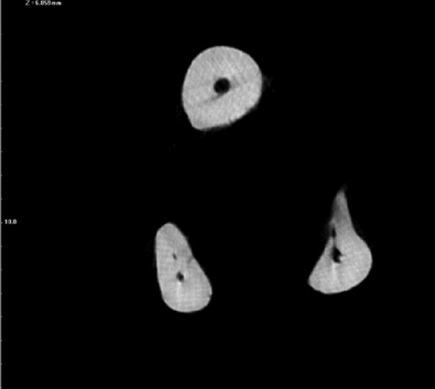
Fig. 2 The accessory canals are seen an axial micro-computed tomography image.
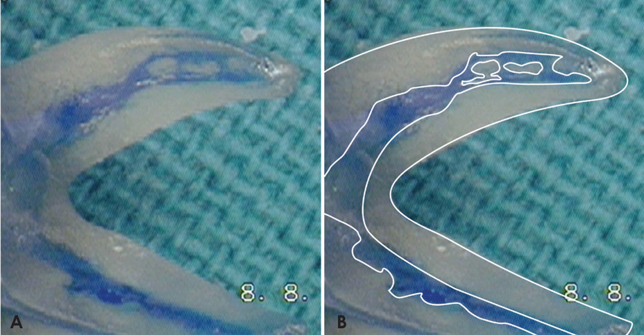
Fig. 3 A. The injected ink is visualized in the accessory canals under the stereomicroscope (10x). B. the main and accessory canals.
Cited by 1 articles
-
Three-dimensional measurement of periodontal surface area for quantifying inflammatory burden
Sa-Beom Park, So-Youn An, Won-Jeong Han, Jong-Tae Park
J Periodontal Implant Sci. 2017;47(3):154-164. doi: 10.5051/jpis.2017.47.3.154.
Reference
-
1. Cantatore G, Berutti E, Castellucci A. Missed anatomy: frequency and clinical impact. Endod Topics. 2006; 15:3–31.
Article2. Schilder H. Cleaning and shaping the root canal. Dent Clin North Am. 1974; 18:269–296.3. Ruddle C. Cleaning and shaping the root canal system. In : Cohen S, Burns RC, editors. Pathways of the pulp. 8th ed. St. Louis: Mosby;2002. p. 231–292.4. Aydin C, Inan U, Yasar S, Bulucu B, Tunca YM. Comparison of shaping ability of RaCe and Hero Shaper instruments in simulated curved canals. Oral Surg Oral Med Oral Pathol Oral Radiol Endod. 2008; 105:e92–e97.
Article5. Bagherian A, Kalhori KA, Sadeghi M, Mirhosseini F, Parisay I. An in vitro study of root and canal morphology of human deciduous molars in an Iranian population. J Oral Sci. 2010; 52:397–403.
Article6. Vertucci FJ, Haddix JE. Chapter 7. Tooth morphology and access cavity preparation. In : Hargreaves KM, Cohen S, editors. Cohen's pathways of the pulp. 10th ed. St. Louis: Mosby;2011. p. 136–222.7. Poornima P, Subba Reddy VV. Comparison of digital radiography, decalcification, and histologic sectioning in the detection of accessory canals in furcation areas of human primary molars. J Indian Soc Pedod Prev Dent. 2008; 26:49–52.
Article8. Fuks AB, Eidelman E, Pauker N. Root fillings with Endoflas in primary teeth: a retrospective study. J Clin Pediatr Dent. 2002; 27:41–45.
Article9. Adorno CG, Yoshioka T, Suda H. Incidence of accessory canals in Japanese anterior maxillary teeth following root canal filling ex vivo. Int Endod J. 2010; 43:370–376.10. Tannure PN, Barcelos R, Portela MB, Gleiser R, Primo LG. Histopathologic and SEM analysis of primary teeth with pulpectomy failure. Oral Surg Oral Med Oral Pathol Oral Radiol Endod. 2009; 108:e29–e33.
Article11. Ringelstein D, Seow WK. The prevalence of furcation foramina in primary molars. Pediatr Dent. 1989; 11:198–202.12. Morabito A, Defabianis P. A SEM investigation on pulpalperiodontal connections in primary teeth. ASDC J Dent Child. 1992; 59:53–57.13. Kumar VD. A scanning electron microscope study of prevalence of accessory canals on the pulpal floor of deciduous molars. J Indian Soc Pedod Prev Dent. 2009; 27:85–89.
Article14. Verma P, Love RM. A micro CT study of the mesiobuccal root canal morphology of the maxillary first molar tooth. Int Endod J. 2011; 44:210–217.
Article15. Scarfe WC, Levin MD, Gane D, Farman AG. Use of cone beam computed tomography in endodontics. Int J Dent. 2009; 2009:634567.
Article16. Kamburoğlu K, Barenboim SF, Arıtürk T, Kaffe I. Quantitative measurements obtained by micro-computed tomography and confocal laser scanning microscopy. Dentomaxillofac Radiol. 2008; 37:385–391.
Article17. Three-dimensional analysis of mesiobuccal root canal of Japanese maxillary first molar using micro-CT. Bull Tokyo Dent Coll. 2011; 52:77–84.18. Somma F, Leoni D, Plotino G, Grande NM, Plasschaert A. Root canal morphology of the mesiobuccal root of maxillary first molars: a micro-computed tomographic analysis. Int Endod J. 2009; 42:165–174.
Article19. Swain MV, Xue J. State of the art of micro-CT applications in dental research. Int J Oral Sci. 2009; 1:177–188.
Article20. Kamburoğlu K, Murat S, Kolsuz E, Kurt H, Yüksel S, Paksoy C. Comparative assessment of subjective image quality of cross-sectional cone-beam computed tomography scans. J Oral Sci. 2011; 53:501–508.
Article21. Garg AK, Tewari RK, Kumar A, Hashmi SH, Agrawal N, Mishra SK. Prevalence of three-rooted mandibular permanent first molars among the Indian population. J Endod. 2010; 36:1302–1306.
Article22. Skidmore AE, Bjorndal AM. Root canal morphology of the human mandibular first molar. Oral Surg Oral Med Oral Pathol. 1971; 32:778–784.
Article23. Neelakantan P, Subbarao C, Subbarao CV, Ravindranath M. Root and canal morphology of mandibular second molars in an Indian population. J Endod. 2010; 36:1319–1322.
Article24. Sperber GH, Moreau JL. Study of the number of roots and canals in Senegalese first permanent mandibular molars. Int Endod J. 1998; 31:117–122.
Article25. Chourasia HR, Meshram GK, Warhadpande M, Dakshindas D. Root canal morphology of mandibular first permanent molars in an Indian population. Int J Dent. 2012; 2012:745152.
Article26. Liu N, Li X, Liu N, Ye L, An J, Nie X, et al. A micro-computed tomography study of the root canal morphology of the mandibular first premolar in a population from southwestern China. Clin Oral Investig. 2013; 17:999–1007.
Article27. Matherne RP, Angelopoulos C, Kulild JC, Tira D. Use of conebeam computed tomography to identify root canal systems in vitro. J Endod. 2008; 34:87–89.
Article28. Blattner TC, George N, Lee CC, Kumar V, Yelton CD. Efficacy of cone-beam computed tomography as a modality to accurately identify the presence of second mesiobuccal canals in maxillary first and second molars: a pilot study. J Endod. 2010; 36:867–870.
Article29. Neelakantan P, Subbarao C, Subbarao CV. Comparative evaluation of modified canal staining and clearing technique, conebeam computed tomography, peripheral quantitative computed tomography, spiral computed tomography, and plain and contrast medium-enhanced digital radiography in studying root canal morphology. J Endod. 2010; 36:1547–1551.
Article30. Wang Y, Zheng QH, Zhou XD, Tang L, Wang Q, Zheng GN, et al. Evaluation of the root and canal morphology of mandibular first permanent molars in a western Chinese population by cone-beam computed tomography. J Endod. 2010; 36:1786–1789.
Article31. Gu L, Wei X, Ling J, Huang X. A microcomputed tomographic study of canal isthmuses in the mesial root of mandibular first molars in a Chinese population. J Endod. 2009; 35:353–356.
Article32. Fan B, Pan Y, Gao Y, Fang F, Wu Q, Gutmann JL. Three-dimensional morphologic analysis of isthmuses in the mesial roots of mandibular molars. J Endod. 2010; 36:1866–1869.
Article33. Baratto Filho F, Zaitter S, Haragushiku GA, de Campos EA, Abuabara A, Correr GM. Analysis of the internal anatomy of maxillary first molars by using different methods. J Endod. 2009; 35:337–342.
Article34. Villas-Bôas MH, Bernardineli N, Cavenago BC, Marciano M, Del Carpio-Perochena A, de Moraes IG, et al. Micro-computed tomography study of the internal anatomy of mesial root canals of mandibular molars. J Endod. 2011; 37:1682–1686.
Article
- Full Text Links
-
- Actions
-
Cited
- CITED
-
- Close
- Share
-
- Similar articles
-
- Analysis of C-shaped root canal configuration in maxillary molars in a Korean population using cone-beam computed tomography
- Isthmuses, accessory canals, and the direction of root curvature in permanent mandibular first molars: an in vivo computed tomography study
- A Study of Root Canals Morphology in Primary Molars using Computerized Tomography
- Assessment of Root and Root Canal Morphology of Human Primary Molars using CBCT
- Comparison of limited- and large-volume cone-beam computed tomography using a small voxel size for detecting isthmuses in mandibular molars
