J Korean Foot Ankle Soc.
2015 Dec;19(4):156-160. 10.14193/jkfas.2015.19.4.156.
Usefulness of Computed Tomography in Patients with Acute Malleolar Fracture
- Affiliations
-
- 1Department of Orthopaedic Surgery, Sanggye Paik Hospital, Inje University College of Medicine, Seoul, Korea. youngos@paik.ac.kr
- 2Department of Orthopaedic Surgery, National Medical Center, Seoul, Korea.
- KMID: 2132457
- DOI: http://doi.org/10.14193/jkfas.2015.19.4.156
Abstract
- PURPOSE
We compared plain radiographs with computed tomography (CT) images to evaluate the usefulness of preoperative CT in acute ankle malleolar fracture in terms of accuracy of diagnosis and planning of operative strategy.
MATERIALS AND METHODS
A retrospective analysis was conducted on 210 cases of malleolar fracture treated at our institute for which plain radiograph and CT were obtained preoperatively. Observers had reviewed plain radiographs and recorded fracture classification, anatomical diagnosis, extent and configuration of fractures and then subsequently reviewed CT images. Records from each image were compared and information regarding the differences in fractures was assessed.
RESULTS
Fractures were notably changed in appearance in 88 cases (41.9%) and diagnosis changed in 30 cases (14.3%). According to the change of diagnosis and fracture appearances, the operative strategy was changed in 15 cases (7.1%) including incision, order of reduction, and target of fixation.
CONCLUSION
CT could be a useful adjunctive imaging tool in addition to the plain radiograph in planning of operative treatment for acute malleolar fracture in terms of estimating exact configuration, extent of fractures and even newly revealed hidden fractures.
Keyword
Figure
-

Figure 1. (A) No definite posterior malleolus fracture was seen in plain radiograph. (B) Posterior malleolar fracture gap was revealed by computed tomography.
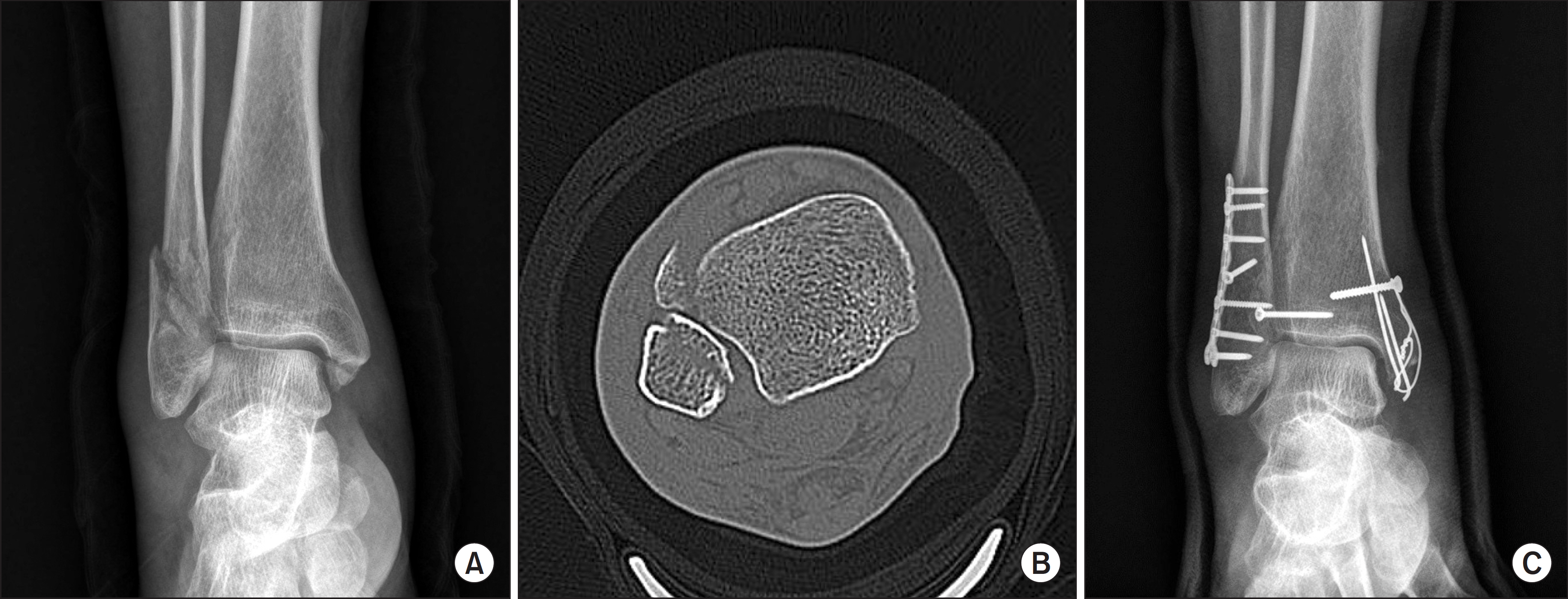
Figure 2. (A) Other fracture line was missed by initial plain radiograph. (B) Additional Chaput tubercle fracture was found after reviewing the axial view of computed tomography image. (C) Chaput tubercle fragment was fixed with one 2.7 mm cortical screw.
Reference
-
References
1. Irwin TA, Lien J, Kadakia AR. Posterior malleolus fracture. J Am Acad Orthop Surg. 2013; 21:32–40.
Article2. Phisitkul P, Ebinger T, Goetz J, Vaseenon T, Marsh JL. Forceps reduction of the syndesmosis in rotational ankle fractures: a cadaveric study. J Bone Joint Surg Am. 2012; 94:2256–61.3. Magid D, Michelson JD, Ney DR, Fishman EK. Adult ankle fractures: comparison of plain films and interactive two- and three-dimensional CT scans. AJR Am J Roentgenol. 1990; 154:1017–23.
Article4. Büchler L, Tannast M, Bonel HM, Weber M. Reliability of radiologic assessment of the fracture anatomy at the posterior tibial plafond in malleolar fractures. J Orthop Trauma. 2009; 23:208–12.
Article5. Ferries JS, DeCoster TA, Firoozbakhsh KK, Garcia JF, Miller RA. Plain radiographic interpretation in trimalleolar ankle fractures poorly assesses posterior fragment size. J Orthop Trauma. 1994; 8:328–31.
Article6. Haraguchi N, Haruyama H, Toga H, Kato F. Pathoanatomy of posterior malleolar fractures of the ankle. J Bone Joint Surg Am. 2006; 88:1085–92.
Article7. Black EM, Antoci V, Lee JT, Weaver MJ, Johnson AH, Susarla SM, et al. Role of preoperative computed tomography scans in operative planning for malleolar ankle fractures. Foot Ankle Int. 2013; 34:697–704.
Article
- Full Text Links
-
- Actions
-
Cited
- CITED
-
- Close
- Share
-
- Similar articles
-
- Usefulness of Computed Tomography on Distal Tibia Intra-Articular Fracture Associated with Spiral Tibia Shaft Fracture
- Assessment of Noncontiguous Posterior Malleolar Fractures in Distal One-Third Tibia Shaft Fractures with Proximal Fibula Fractures
- Comparison of the Size of the Posterior Malleolar Fragment in Trimalleolar Ankle Fractures Measured Using Lateral Plain Radiography and Three-Dimensional Computed Tomography
- The Clinical Study of Ankle Fracture and Dislocation
- HERBERT SCREW FIXATION FOR NON-COMMINUTED CLOSED MEDIAL MALLEOLAR FRACTURE
