Integrated three-dimensional digital assessment of accuracy of anterior tooth movement using clear aligners
- Affiliations
-
- 1Department of Orthodontics, Capital Medical University School of Stomatology, Beijing, China. hongmingguocn@163.com
- 2Department of Orthodontics, Luhe Hospital of China Capital Medical University, Beijing, China.
- 3Department of Orthodontics, Yan'an University Affiliated Hospital, Yan'an, China.
- 4Private Practice, Beijing, China.
- KMID: 2130589
- DOI: http://doi.org/10.4041/kjod.2015.45.6.275
Abstract
OBJECTIVE
To assess the accuracy of anterior tooth movement using clear aligners in integrated three-dimensional digital models.
METHODS
Cone-beam computed tomography was performed before and after treatment with clear aligners in 32 patients. Plaster casts were laser-scanned for virtual setup and aligner fabrication. Differences in predicted and achieved root and crown positions of anterior teeth were compared on superimposed maxillofacial digital images and virtual models and analyzed by Student's t-test.
RESULTS
The mean discrepancies in maxillary and mandibular crown positions were 0.376 +/- 0.041 mm and 0.398 +/- 0.037 mm, respectively. Maxillary and mandibular root positions differed by 2.062 +/- 0.128 mm and 1.941 +/- 0.154 mm, respectively.
CONCLUSIONS
Crowns but not roots of anterior teeth can be moved to designated positions using clear aligners, because these appliances cause tooth movement by tilting motion.
Figure
-
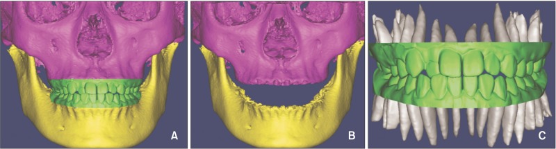
Figure 1 Construction of the integrated three-dimensional digital model. A, The integrated model; B, images of the maxilla and mandible; C, images of the complete dentition.
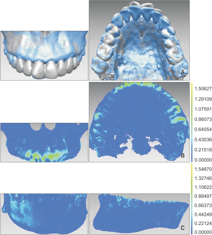
Figure 2 Pretreatment and post-treatment registration of the jaws. A, Maxillary registration. B, Mandibular registration. Blue indicates post-treatment and silver indicates pretreatment. C, Detection map after registration. Dark blue is visible (registration accuracy ≤ 0.15 mm).
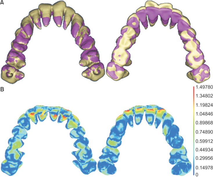
Figure 3 Cone-beam computed tomography-based registration of crown and root positions. A, Comparison of pretreatment (yellow) and post-treatment (pink) root positions. A small amount of movement is visible in the apical part while the coronal part appears to have moved to a great extent. B, Detection map after registration. The crown and most of the apical part appear dark blue (registration accuracy ≤ 0.15 mm), while the anterior crown is red.
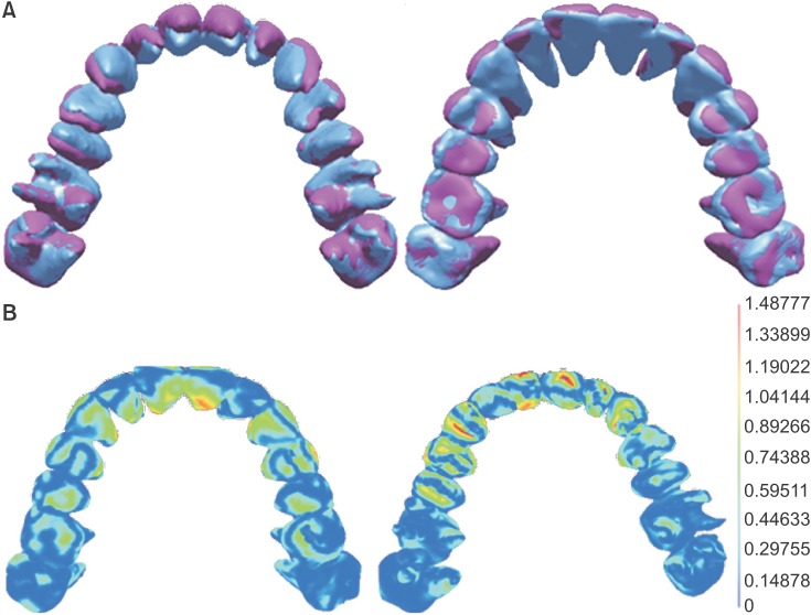
Figure 4 Discrepancies in crown and root positions after treatment with clear aligners. A, Comparison of achieved (blue) and predicted (pink) crown and root positions. Only the crown reached the predicted position. B, Detection map after registration. The immovable and moved parts of molar teeth appear dark blue (registration accuracy ≤ 0.15 mm), while the crown appears red.
Cited by 2 articles
-
A systematic review of the accuracy and efficiency of dental movements with Invisalign®
Lidia Galan-Lopez, Jorge Barcia-Gonzalez, Eliseo Plasencia
Korean J Orthod. 2019;49(3):140-149. doi: 10.4041/kjod.2019.49.3.140.A comparison of the precision of three-dimensional images acquired by 2 digital intraoral scanners: effects of tooth irregularity and scanning direction
Ji-won Anh, Ji-Man Park, Youn-Sic Chun, Miae Kim, Minji Kim
Korean J Orthod. 2016;46(1):3-12. doi: 10.4041/kjod.2016.46.1.3.
Reference
-
1. Kassas W, Al-Jewair T, Preston CB, Tabbaa S. Assessment of Invisalign treatment outcomes using the ABO Model Grading System. J World Fed Orthod. 2013; 2:e61–e64.
Article2. Kravitz ND, Kusnoto B, BeGole E, Obrez A, Agran B. How well does Invisalign work? A prospective clinical study evaluating the efficacy of tooth movement with Invisalign. Am J Orthod Dentofacial Orthop. 2009; 135:27–35. PMID: 19121497.
Article3. Lund H, Gröndahl K, Hansen K, Gröndahl HG. Apical root resorption during orthodontic treatment. A prospective study using cone beam CT. Angle Orthod. 2012; 82:480–487. PMID: 21919826.4. Krieger E, Seiferth J, Marinello I, Jung BA, Wriedt S, Jacobs C, et al. Invisalign® treatment in the anterior region: were the predicted tooth movements achieved? J Orofac Orthop. 2012; 73:365–376. PMID: 22890691.5. Castroflorio T, Garino F, Lazzaro A, Debernardi C. Upper-incisor root control with Invisalign appliances. J Clin Orthod. 2013; 47:346–351. PMID: 23863556.6. Guo H, Zhou J, Bai Y, Li S. A three-dimensional setup model with dental roots. J Clin Orthod. 2011; 45:209–216. PMID: 21785205.7. Chisari JR, McGorray SP, Nair M, Wheeler TT. Variables affecting orthodontic tooth movement with clear aligners. Am J Orthod Dentofacial Orthop. 2014; 145(4 Suppl):S82–S91. PMID: 24680028.
Article8. Rossini G, Parrini S, Castroflorio T, Deregibus A, Debernardi CL. Efficacy of clear aligners in controlling orthodontic tooth movement: A systematic review. Angle Orthod. 2015; 85:881–889. PMID: 25412265.
Article9. Djeu G, Shelton C, Maganzini A. Outcome assessment of Invisalign and traditional orthodontic treatment compared with the American Board of Orthodontics objective grading system. Am J Orthod Dentofacial Orthop. 2005; 128:292–298. PMID: 16168325.
Article10. Nguyen CV, Chen J. Chapter 14. In : Tuncay OC, editor. The invisalign system. London, UK: Quintessence Publishing Company, Ltd.;2006. p. 12–32.11. Kim DS, Choi SC, Lee SS, Heo MS, Huh KH, Hwang SJ, et al. Principal direction of inertia for 3D trajectories from patient-specific TMJ movement. Comput Biol Med. 2013; 43:169–175. PMID: 23321156.
Article12. Macchi A, Carrafiello G, Cacciafesta V, Norcini A. Three-dimensional digital modeling and setup. Am J Orthod Dentofacial Orthop. 2006; 129:605–610. PMID: 16679200.
Article13. Kim BC, Lee CE, Park W, Kang SH, Zhengguo P, Yi CK, et al. Integration accuracy of digital dental models and 3-dimensional computerized tomography images by sequential point- and surface-based markerless registration. Oral Surg Oral Med Oral Pathol Oral Radiol Endod. 2010; 110:370–378. PMID: 20591700.
Article14. Ye N, Jian F, Xue J, Wang S, Liao L, Huang W, et al. Accuracy of in-vitro tooth volumetric measurements from cone-beam computed tomography. Am J Orthod Dentofacial Orthop. 2012; 142:879–887. PMID: 23195374.
Article15. Miller RJ, Kuo E, Choi W. Validation of Align Technology's Treat III digital model superimposition tool and its case application. Orthod Craniofac Res. 2003; 6(Suppl 1):143–149. PMID: 14606547.16. Hahn W, Zapf A, Dathe H, Fialka-Fricke J, Fricke-Zech S, Gruber R, et al. Torquing an upper central incisor with aligners--acting forces and biomechanical principles. Eur J Orthod. 2010; 32:607–613. PMID: 20462912.
Article17. Brezniak N. The clear plastic appliance: a biomechanical point of view. Angle Orthod. 2008; 78:381–382. PMID: 18251593.18. Baldwin DK, King G, Ramsay DS, Huang G, Bollen AM. Activation time and material stiffness of sequential removable orthodontic appliances. Part 3: premolar extraction patients. Am J Orthod Dentofacial Orthop. 2008; 133:837–845. PMID: 18538247.
Article19. Krieger E, Drechsler T, Schmidtmann I, Jacobs C, Haag S, Wehrbein H. Apical root resorption during orthodontic treatment with aligners? A retrospective radiometric study. Head Face Med. 2013; 9:21. PMID: 23941626.
Article20. Weltman B, Vig KW, Fields HW, Shanker S, Kaizar EE. Root resorption associated with orthodontic tooth movement: a systematic review. Am J Orthod Dentofacial Orthop. 2010; 137:462–476. PMID: 20362905.
Article21. Apajalahti S, Peltola JS. Apical root resorption after orthodontic treatment -- a retrospective study. Eur J Orthod. 2007; 29:408–412. PMID: 17631606.
- Full Text Links
-
- Actions
-
Cited
- CITED
-
- Close
- Share
-
- Similar articles
-
- Accuracy of orthodontic movements with 3D printed aligners: A prospective observational pilot study
- Achievement of Anterior Teeth Arrangement Using a Single Stage of Clear Aligner
- Force changes associated with differential activation of en-masse retraction and/or intrusion with clear aligners
- Clear Aligner Therapy: Evidence, Eloquence and Reality
- Force Assessment of Thermoformed and Direct-printed Aligners in a Lingual Bodily Movement of a Central Incisor Over Time: A 14-day In Vitro Study
