J Gynecol Oncol.
2008 Jun;19(2):108-112. 10.3802/jgo.2008.19.2.108.
Value of pelvic examination and imaging modality for the evaluation of tumor size in cervical cancer
- Affiliations
-
- 1Department of Obstetrics and Gynecology, Seoul National University, College of Medicine, Seoul, Korea. kjwksh@snu.ac.kr
- 2Department of Obstetrics and Gynecology, College of Medicine, Chung-Ang University, Seoul, Korea.
- KMID: 2129909
- DOI: http://doi.org/10.3802/jgo.2008.19.2.108
Abstract
-
OBJECTIVE
The purpose of this study was to compare the accuracy of pelvic examination versus imaging modality such as computed tomography (CT) or magnetic resonance imaging (MRI) in the measurement of the tumor size of invasive cervical carcinoma based on pathologic findings.
METHODS
Patients with stage Ib-II cervical cancer who underwent primary surgical treatment between January 2003 and December 2005 were evaluated retrospectively. One hundred three consecutive patients aged 24 to 81 years (mean age, 50.6 years), who had not received any treatment previously were included in this study. Accuracy of preoperative CT or MRI versus pelvic examination in the measurement of tumor size was compared based on pathologic findings. All patients were examined and staged clinically by the gynecologic oncologist. Surgery was performed within 2 weeks after imaging studies. The data were analyzed using descriptive statistics.
RESULTS
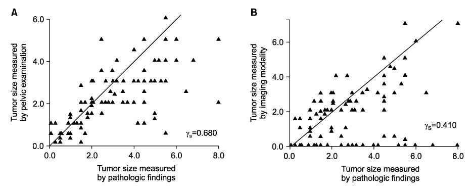
The largest diameter of the tumor measured by pathologic findings was 2.76+/-1.76 cm. Based on pathologic findings, accuracy was estimated by the degree of agreement with a difference of <0.5 or 1.0 cm between the measurements of tumor size obtained by pelvic examination and imaging modality. Pelvic examination and imaging modality had an accuracy of 46.6% and 39.8%, respectively, with a difference of <0.5 cm, and an accuracy of 72.8% and 55.3%, respectively, with a difference of <1.0 cm. Correlation with pathologic findings was higher for pelvic examination (r(s)=0.680) than for imaging modality (r(s)=0.410). In determining the size of tumor mass differentiating >4.0 cm from < or =4.0 cm, imaging modality showed higher accuracy than pelvic examination.
CONCLUSION
For the patients with stage Ib to II cervical cancer, pelvic examination is superior to imaging modality with regard to evaluation of the tumor size. However, imaging modality may be accurate for evaluating bulky tumors of cervical cancer.
Keyword
MeSH Terms
Figure
Reference
-
1. Korean Society of Obstetrics and Gynecology, Committee of Gynecologic Oncology. Annual report of gynecologic cancer registry program in Korea for 2004 (2004.1.1-2004.12.31). Korean J Obstet Gynecol. 2007. 50:28–78.2. Lai CH, Hong JH, Hsueh S, Ng KK, Chang TC, Tseng CJ, et al. Preoperative prognostic variables and the impact of postoperative adjuvant therapy on the outcomes of Stage IB or II cervical carcinoma patients with or without pelvic lymph node metastases: An analysis of 891 cases. Cancer. 1999. 85:1537–1546.3. Sedlis A, Bundy BN, Rotman MZ, Lentz SS, Muderspach LI, Zaino RJ. A randomized trial of pelvic radiation therapy versus no further therapy in selected patients with stage IB carcinoma of the cervix after radical hysterectomy and pelvic lymphadenectomy: A Gynecologic Oncology Group Study. Gynecol Oncol. 1999. 73:177–183.4. Zaino RJ, Ward S, Delgado G, Bundy B, Gore H, Fetter G, et al. Histopathologic predictors of the behavior of surgically treated stage IB squamous cell carcinoma of the cervix. A Gynecologic Oncology Group study. Cancer. 1992. 69:1750–1758.5. Vinh-Hung V, Bourgain C, Vlastos G, Cserni G, De Ridder M, Storme G, et al. Prognostic value of histopathology and trends in cervical cancer: A SEER population study. BMC Cancer. 2007. 7:164.6. Metindir J, Bilir G. Prognostic factors affecting disease-free survival in early-stage cervical cancer patients undergoing radical hysterectomy and pelvic-paraaortic lymphadenectomy. Eur J Gynaecol Oncol. 2007. 28:28–32.7. Chung CK, Nahhas WA, Stryker JA, Curry SL, Abt AB, Mortel R. Analysis of factors contributing to treatment failures in stages IB and IIA carcinoma of the cervix. Am J Obstet Gynecol. 1980. 138:550–556.8. Homesley HD, Raben M, Blake DD, Ferree CR, Bullock MS, Linton EB, et al. Relationship of lesion size to survival in patients with stage IB squamous cell carcinoma of the cervix uteri treated by radiation therapy. Surg Gynecol Obstet. 1980. 150:529–531.9. Kristensen GB, Abeler VM, Risberg B, Trop C, Bryne M. Tumor size, depth of invasion, and grading of the invasive tumor front are the main prognostic factors in early squamous cell cervical carcinoma. Gynecol Oncol. 1999. 74:245–251.10. Trattner M, Graf AH, Lax S, Forstner R, Dandachi N, Haas J, et al. Prognostic factors in surgically treated stage ib-iib cervical carcinomas with special emphasis on the importance of tumor volume. Gynecol Oncol. 2001. 82:11–16.11. Horn LC, Fischer U, Raptis G, Bilek K, Hentschel B. Tumor size is of prognostic value in surgically treated FIGO stage II cervical cancer. Gynecol Oncol. 2007. 107:310–315.12. Choi HJ, Kim SH, Seo SS, Kang S, Lee S, Kim JY, et al. MRI for pretreatment lymph node staging in uterine cervical cancer. AJR Am J Roentgenol. 2006. 187:W538–W543.13. Soutter WP, Hanoch J, D'Arcy T, Dina R, McIndoe GA, DeSouza NM. Pretreatment tumour volume measurement on high-resolution magnetic resonance imaging as a predictor of survival in cervical cancer. BJOG. 2004. 111:741–747.14. Arimoto T. Significance of computed tomography-measured volume in the prognosis of cervical carcinoma. Cancer. 1993. 72:2383–2388.15. Botsis D, Gregoriou O, Kalovidouris A, Tsarouchis K, Zourlas PA. The value of computed tomography in staging cervical carcinoma. Int J Gynaecol Obstet. 1988. 27:213–218.16. Camilien L, Gordon D, Fruchter RG, Maiman M, Boyce JG. Predictive value of computerized tomography in the presurgical evaluation of primary carcinoma of the cervix. Gynecol Oncol. 1988. 30:209–215.17. Hoffman MS, Cardosi RJ, Roberts WS, Fiorica JV, Grendys EC Jr, Griffin D. Accuracy of pelvic examination in the assessment of patients with operable cervical cancer. Am J Obstet Gynecol. 2004. 190:986–993.18. Alvarez RD, Potter ME, Soong SJ, Gay FL, Hatch KD, Partridge EE, et al. Rationale for using pathologic tumor dimensions and nodal status to subclassify surgically treated stage IB cervical cancer patients. Gynecol Oncol. 1991. 43:108–112.19. Averette HE, Ford JH Jr, Dudan RC, Girtanner RE, Hoskins WJ, Lutz MH. Staging of cervical cancer. Clin Obstet Gynecol. 1975. 18:215–232.20. Van Nagell JR Jr, Roddick JW Jr, Lowin DM. The staging of cervical cancer: Inevitable discrepancies between clinical staging and pathologic findinges. Am J Obstet Gynecol. 1971. 110:973–978.21. Hricak H, Lacey CG, Sandles LG, Chang YC, Winkler ML, Stern JL. Invasive cervical carcinoma: Comparison of MR imaging and surgical findings. Radiology. 1988. 166:623–631.22. Mayr NA, Yuh WT, Zheng J, Ehrhardt JC, Sorosky JI, Magnotta VA, et al. Tumor size evaluated by pelvic examination compared with 3-D quantitative analysis in the prediction of outcome for cervical cancer. Int J Radiat Oncol Biol Phys. 1997. 39:395–404.23. Sironi S, De Cobelli F, Scarfone G, Colombo E, Bolis G, Ferrari A, et al. Carcinoma of the cervix: Value of plain and gadolinium-enhanced MR imaging in assessing degree of invasiveness. Radiology. 1993. 188:797–801.24. Kim SH, Choi BI, Han JK, Kim HD, Lee HP, Kang SB, et al. Preoperative staging of uterine cervical carcinoma: Comparison of CT and MRI in 99 patients. J Comput Assist Tomogr. 1993. 17:633–640.25. Togashi K, Nishimura K, Sagoh T, Minami S, Noma S, Fujisawa I, et al. Carcinoma of the cervix: Staging with MR imaging. Radiology. 1989. 171:245–251.26. Hricak H, Gatsonis C, Chi DS, Amendola MA, Brandt K, Schwartz LH, et al. Role of imaging in pretreatment evaluation of early invasive cervical cancer: Results of the intergroup study American College of Radiology Imaging Network 6651-Gynecologic Oncology Group 183. J Clin Oncol. 2005. 23:9329–9337.
- Full Text Links
- Actions
-
Cited
- CITED
-
- Close
- Share
- Similar articles
-
- Predictive Value of Clinical Examination, Computed Tomography and Magnetic Resonance Imaging in the Clinical Staging of the Cervical Carcinoma
- Treatment Planning Correction Using MRI in the Radiotherapy of Cervical Cancer
- Staging of uterine cervical carcinoma: comparison of CT and MR imaging
- Differences of Tumor Size Measured by Ultrasonography and Magnetic Resonance Imaging Compared to Pathological Tumor Size in Primary Breast Cancer
- Estimation by Gross Findings in Early Gastric Cancer


