A Case of Imploding Antrum (Silent Sinus) Syndrome after Orbital Decompression
- Affiliations
-
- 1Department of Ophthalmology, Samsung Medical Center, Sungkyunkwan University School of Medicine, Seoul, Korea. ydkimoph@skku.edu
- 2Department of Ophthalmology, Korea Veterans Hospital, Seoul, Korea.
- KMID: 2127130
- DOI: http://doi.org/10.3341/jkos.2008.49.2.362
Abstract
-
PURPOSE: Imploding antrum (silent sinus) syndrome has clinical features of enophthalmos and hypoglobus after a downward collapse of inferior orbital wall with an ipsilateral volume decrease of maxillary sinus. We present a case of imploding antrum syndrome after an orbital decompression surgery.
CASE SUMMARY
A 26-year-old female underwent inferomedial wall orbital decompression surgery through a caruncular approach to reduce exophthalmos. At 14 months after surgery, her right eye showed 2 mm of enophthalmos and orbital CT scan revealed both maxillary sinusitis. At 28 months after surgery, 3 mm of enophthalmos and hypoglobus of the right were observed, and an orbital CT scan was taken. Orbital CT scan showed a downward collapse of inferior orbital wall, a volume decrease and inward bowing of the maxillary sinus, and a maxillary opacification on the right side, which are typical findings of imploding antrum (silent sinus) syndrome.
CONCLUSIONS
Imploding antrum (silent sinus) syndrome after orbital decompression surgery is a rare complication. Considering that any prolapsed orbital fat after orbital decompression surgery could result in imploding antrum syndrome with hypoventilation of a maxillary sinus, care should be taken to keep the maxillo-ethmoidal interface (bony strut) intact, which helps maintain maxillary aeration.
MeSH Terms
Figure
-

Figure 1. A 26-year-old woman with thyroid orbitopathy showed bilateral exophthalmos and ptosis in the left eye before orbital decompression.

Figure 2. A facial photograph (A) taken at 14 months after orbital decompression surgery showed 2 mm of enophthalmos in the right eye. Axial (B) and coronal (C) CT scans showed enophthalmos of the right eye, bilateral maxillary sinusitis. The medial and inferior orbital walls were properly decompressed.
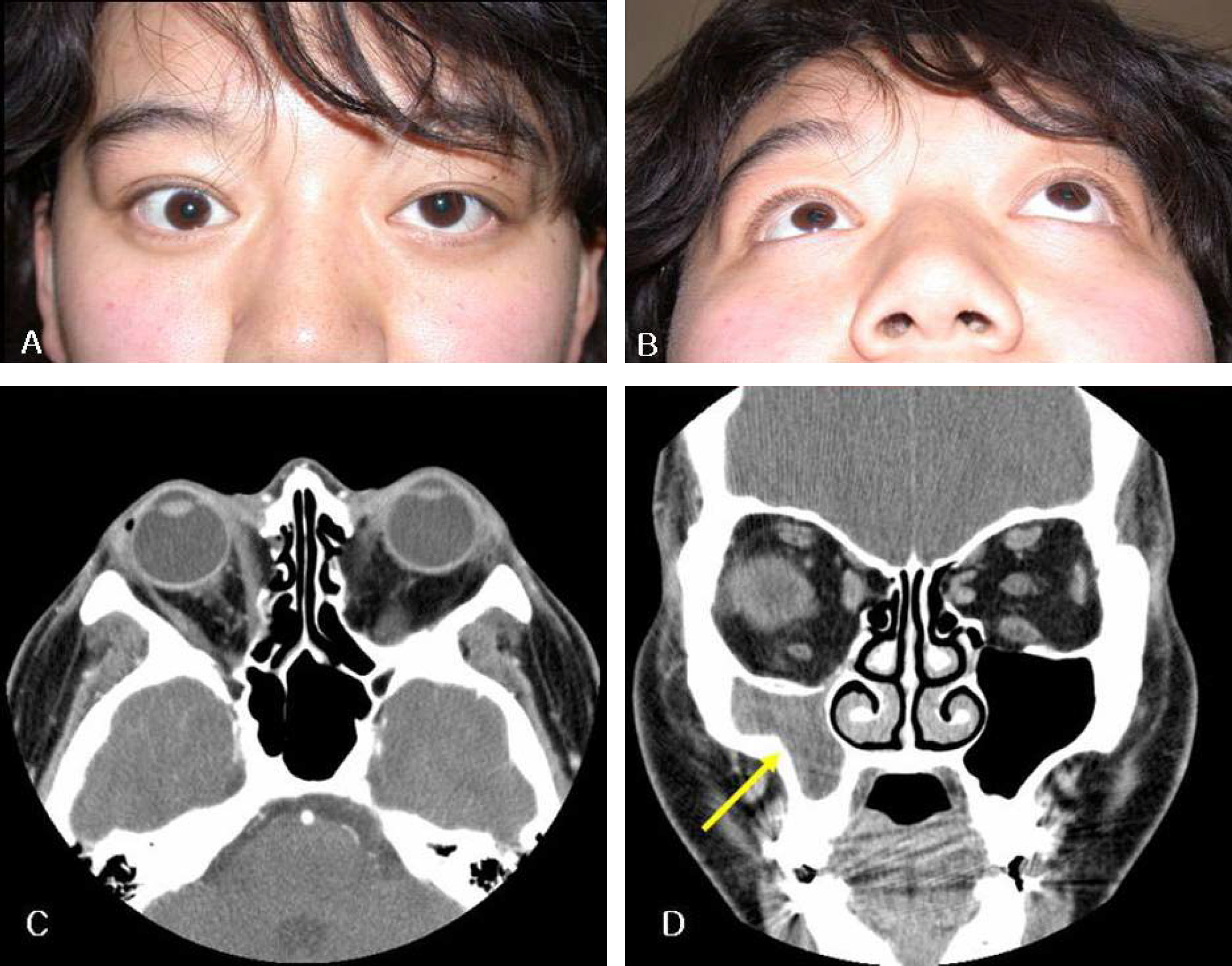
Figure 3. Facial photographs (A, B) taken at 28 months after bilateral orbital decompression showed 2 mm of hypoglobus and 3 mm of enophthalmos in the right eye. Axial (C) and coronal (D) orbital CT scans demonstrated enophthalmos in the right eye, right maxillary sinus smaller than the left, inferiorly collapsed roof of the right maxillary sinus, and marked inward bowing (implosion) of the lateral wall of the maxilla (arrow).

Figure 4. Facial photographs (A, B) taken at 14 months after inferior meatal antrostomy showed 1.3 mm of hypoglobus and 1 mm of enophthalmos in the right eye. Axial orbital CT scan (C) demonstrated improved enophthalmos in the right eye, compared with Fig. 3C. Coronal orbital CT scan (D) revealed the expansion of the right maxillary sinus cavity after inferior meatal antrostomy, which was evident by the decreased difference in size of the right and left maxillary sinus, compared with Fig. 3D. Mucopurulent material in the maxillary cavity shown in Fig. 3D disappeared completely.
Reference
-
References
1. Mourits MP, Koornneef L, Wiersinga WM, et al. Orbital decompression for Graves' ophthalmopathy by inferomedial, by inferomedial plus lateral, and by coronal approach. Ophthalmology. 1990; 97:636–41.
Article2. Lyons CJ, Rootman J. Orbital decompression for disfiguring exophthalmos in thyroid orbitopathy. Ophthalmology. 1994; 101:223–30.
Article3. Fatourechi V, Garrity JA, Bartley GB, et al. Graves ophthalmopathy. Results of transantral orbital decompression performed primarily for cosmetic indications. Ophthalmology. 1994; 101:938–42.4. Kalmann R, Mourits MP, van der Pol JP, Koornneef L. Coronal approach for rehabilitative orbital decompression in Graves' ophthalmopathy. Br J Ophthalmol. 1997; 81:41–5.
Article5. Rose GE, Lund VJ. Clinical Features and Treatment of Late Enophthalmos after Orbital Decompression: a condition suggesting cause for idiopathic “imploding antrum” (silent sinus) syndrome. Ophthalmology. 2003; 110:819–26.6. Soparkar CN, Patrinely JR, Cuaycong MJ, et al. The silent sinus syndrome. A cause of spontaneous enophthalmos. Ophthalmology. 1994; 101:772–8.7. Rose GE, Sandy CJ, Halberg L, Moseley I. Clinical and radiological characteristics of the imploding antrum, or “silent sinus,” syndrome. Ophthalmology. 2003; 110:811–8.8. Cline RA, Rootman J. Enophthalmos: a clinical review. Ophthalmology. 1984; 91:229–37.9. Rodriguez ED, Langner RB, Manson PN. Microsurgical Enophthalmos Correction After Silent Sinus Syndrome. J Craniofac Surg. 2007; 18:454–6.
Article10. Hunt SM, Tami TA. Sinusitis-induced enophthalmos: the silent sinus syndrome. Ear Nose Throat J. 2000; 79:576. 579-81, 584.
Article11. Gillman GS, Schaitkin BM, May M. Asymptomatic enophthalmos: the silent sinus syndrome. Am J Rhinology. 1999; 13:459–62.
Article12. Audemard D, Galipienzo V, Marck E, et al. Silent sinus syndrome: a rare case of enophthalmia. J Fr Ophtalmol. 2002; 25:266–9.13. Gagnon MR, Yeatts RP, Williams Z, Matthews B. Delayed enophthalmos following a minimally displaced orbital floor fracture. Ophthal Plast Reconstr Surg. 2004; 20:241–3.
Article14. Hobbs CGL, Saunders MW, Potts MJ. “Imploding antrum” or silent sinus syndrome following naso-tracheal intubation. Br J Ophthalmol. 2004; 88:974–5.
Article15. Davidson JK, Soparkar CN, Williams JB, Patrinely JR. Negative sinus pressure and normal predisease imaging in silent sinus syndrome. Arch Ophthalmol. 1999; 117:1653–4.16. Ando A, Velasco AA. Management of enophthalmos and superior sulcus deformity induced by the silent sinus syndrome. Aesthetic Plast Surg. 2005; 29:74–7.
Article17. Goldberg RA, Shorr N, Cohen MS. The medial orbital strut in the prevention of postdecompression dystopia in dysthyroid ophthalmopathy. Ophthal Plast Reconstr Surg. 1992; 8:32–4.
Article
- Full Text Links
-
- Actions
-
Cited
- CITED
-
- Close
- Share
-
- Similar articles
-
- A Case of Orbital Apex Syndrome Caused by Mucocele in the Sphenoid Sinus
- Endoscopic Orbital Decompression for Dysthyroid Orbitopathy
- Traumatic Superior orbital fissure syndrome complicating fractures of the facial skeleton: Report of a Case
- Effect of Posterior Strut Removal during Orbital Decompression for Graves’ Orbitopathy
- Change in Quality of Life after Orbital Decompression Surgery in Patients with Dysthyroid Ophthalmopathy
