Comparison of Long-Term Results between One-piece and Three-piece Acrylate Intraocular Lens
- Affiliations
-
- 1Department of Ophthalmology, Chonnam National University Medical School & Hospital, Chonnam National Research Institute for Medical Sciences, Gwangju, Korea. kcyoon@chonnam.ac.kr
- KMID: 2127115
- DOI: http://doi.org/10.3341/jkos.2008.49.2.245
Abstract
-
PURPOSE: To compare the long-term clinical results of one-piece Acrysof(R) (SA60AT) hydrophobic acrylic intraocular lens (IOL) implantation compared with implantation of three-piece Acrysof(R) (MA60BM) hydrophobic acrylic IOL.
METHODS
We retrospectively analyzed each 50 eyes of 50 patients underwent MA60BM or SA60AT IOL implantation and followed for at least 6 months.
RESULTS
Final visual acuity of 0.5 or better was 38 eyes (76%) and 0.8 or better was 20 eyes (40%) in the SA60AT group. In the MA60BM group, it was 41 eyes (82%) and 23 eyes (46%) respectively. There were no significantly differences in predictability, intraocular pressure, endothelial cell density, astigmatism, and incidence of posterior capsule opacification between the two groups. Spherical equivalent at postoperative 1 week was -0.50+/-0.95D in SA60AT group and was -0.31+/-0.88D in MA60BM group (P=0.04). However, there was no significant difference between the two groups during follow up period. In MA60BM group, anterior chamber depth (P=0.02) and distance between iris and IOL (P=0.04) reduced significantly during the first postoperative month.
CONCLUSIONS
Early postoperative axial displacement and changes in spherical equivalent can be occurred in MA60BM group. However there was no difference in long-term clinical results between SA60AT and MA60BM groups.
MeSH Terms
Figure
-
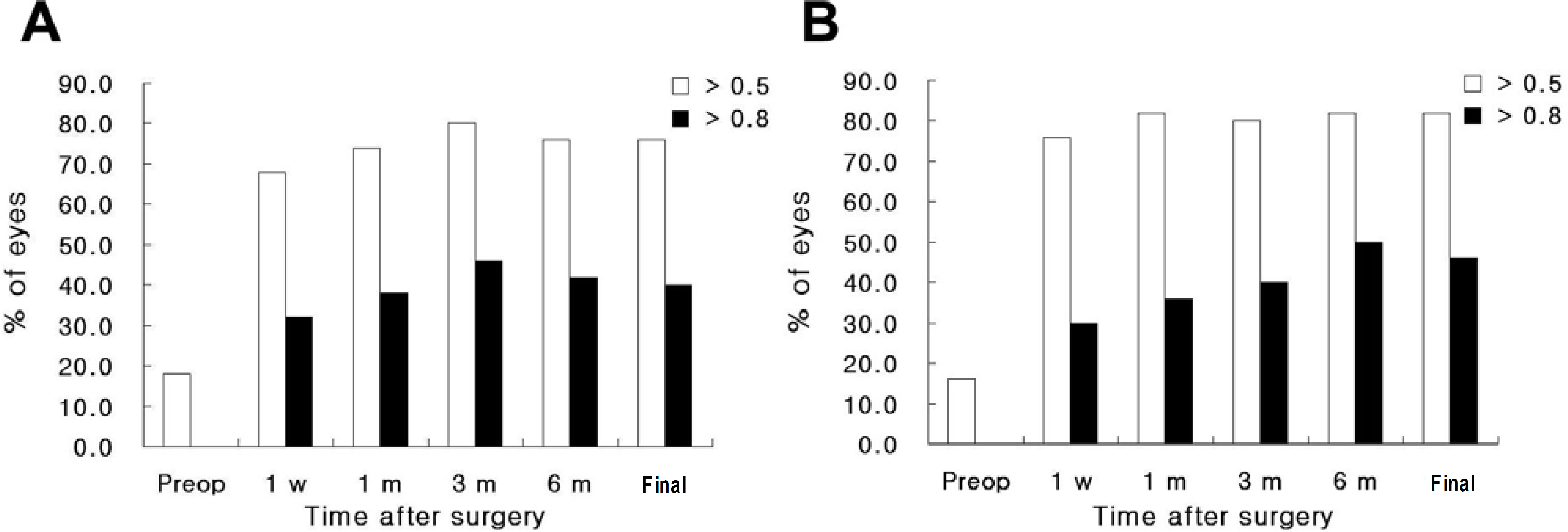
Figure 1. Changes of preoperative and postoperative uncorrected visual acuity between SA60AT (A) and MA60BM (B) groups.

Figure 2. Changes of preoperative and postoperative best corrected visual acuity between SA60AT (A) and MA60BM (B) groups.
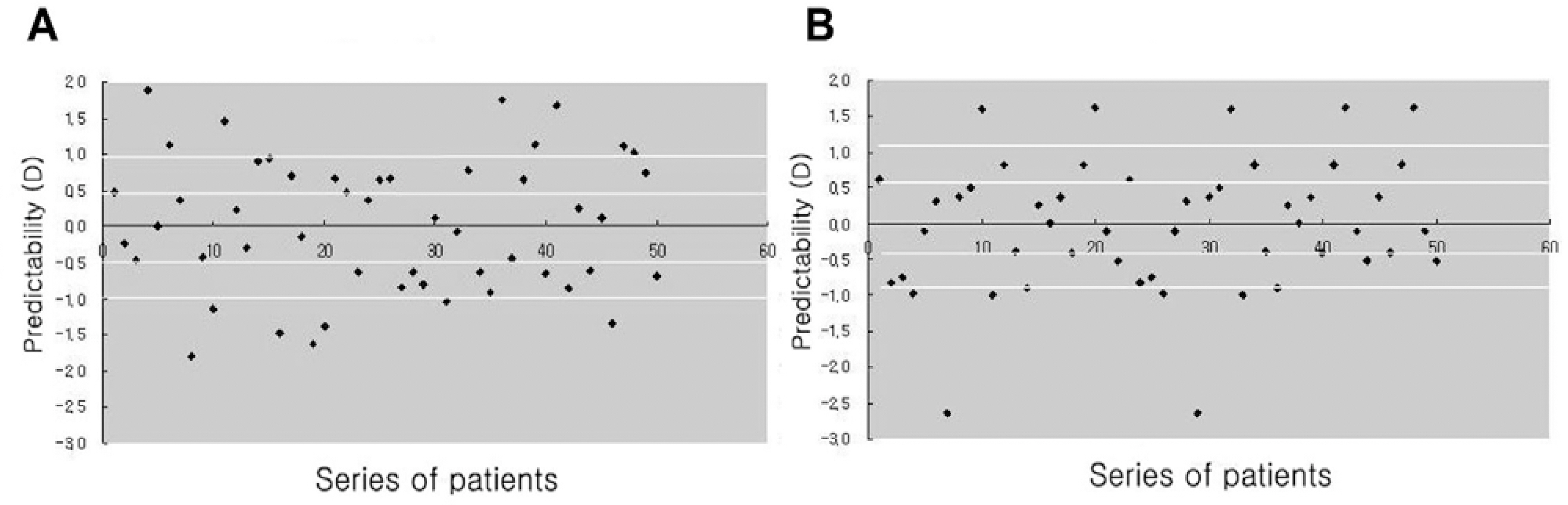
Figure 3. Predictability of the manifest refraction with SA60AT (A) and MA60BM (B) at the final follow-up.
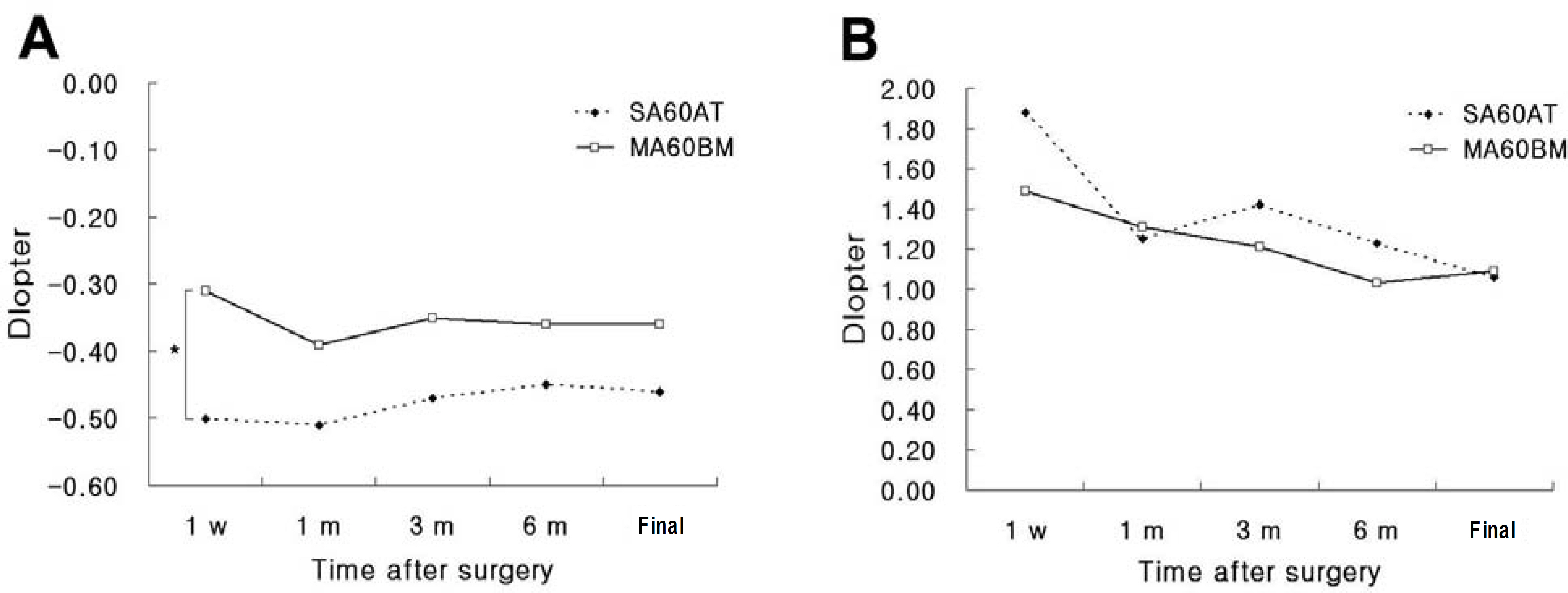
Figure 4. Changes of the postoperative spherical equivalent (A) and astigmatism (B) (*: P<0.05).
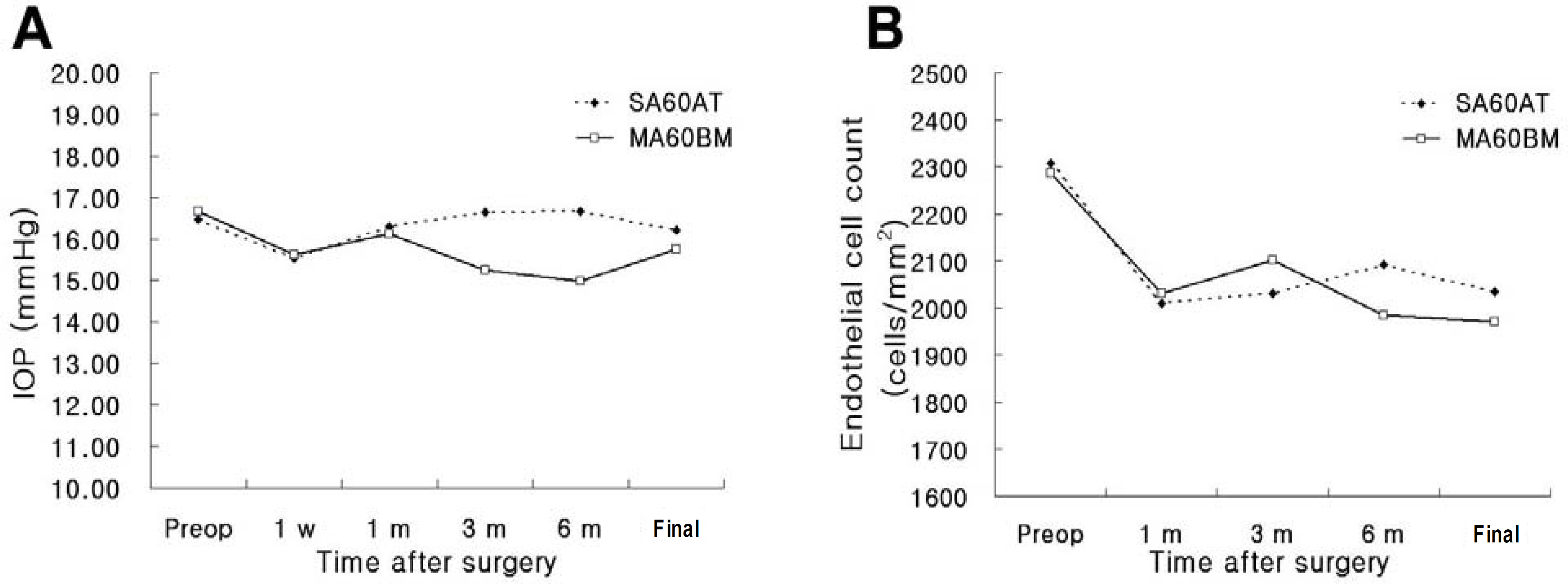
Figure 5. Changes of the postoperative intraocular pressure (A) and endothelial cell count (B).
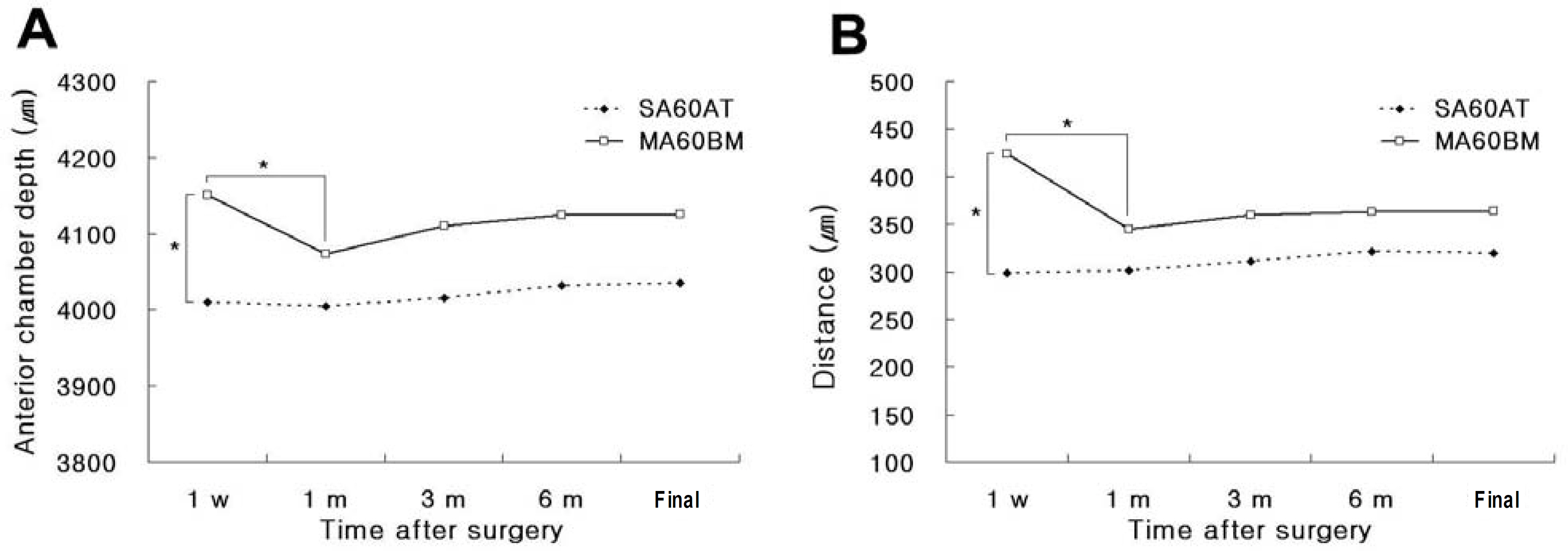
Figure 6. Changes of the postoperative anterior chamber depth (A) and the distance between posterior surface of iris and anterior surface of intraocular lens (B) (*: P<0.05).
Cited by 1 articles
-
Comparison of Visual Function Among Aspheric Intraocular Lenses
In Seong Kang, In Cheon You, Yeoung Geol Park, Kyung Chul Yoon
J Korean Ophthalmol Soc. 2009;50(5):691-697. doi: 10.3341/jkos.2009.50.5.691.
Reference
-
References
1. Kohnen S, Ferrer A, Brauweiler P. Visual function in pseudophakic eyes with poly (methyl methacrylate), silicone, and acrylic intraocular lenses. J Cataract Refract Surg. 1996; 22:1303–7.2. Hayashi K, Hayashi H, Nakao F, Fumihiko H. Reduction in the area of the anterior capsule opening after poltmethymethacrylate, silicone, and soft acrylic intraocular lens implantation. Am J Ophthalmol. 1997; 123:441–7.3. Magual E, Garcia J, Elvira JC, Hueso JR. Clinical results of AcrySof intraocular lens implantation. J Cataract Refract Surg. 1998; 24:114–7.4. Oshika T, Suzuki Y, Kizaki H, Yaguchi S. Two year clinical study of a soft acrylic intraocular lens. J Cataract Refract Surg. 1996; 22:104–9.
Article5. Ursell PG, Spalton DJ, Pande MV, et al. Relationship between intraocular lens biomaterials and posterior capsule opacification. J Cataract Refract Surg. 1998; 24:352–6.
Article6. Cha YD, Oh SH, Lee DH. Comparative assessment of clinical results in various acrylate IOLs. J Korean Ophthalmol Soc. 2006; 47:740–7.7. Choi JA, Kim TI, Tchah HW. Aberration change in pseudophakia with three types of acryl intraocular lens. J Korean Ophthalmol Soc. 2004; 45:405–12.8. Oh SH, Kim JK, Lee DH. The clinical results of hydrophobic single-piece acrylic intraocular lenses after cataract surgery. J Korean Ophthalmol Soc. 2004; 45:2007–13.9. Seo JH, Kim KB, Seo JB. A clinical comparison of Acrysof with hydrophilic acrylic IOLs. J Korean Ophthalmol Soc. 2001; 42:266–71.10. Nishi O, Nishi K. Effect of the optic size of a single-piece acrylic intraocular lens on posterior capsule opacification. J Cataract Refract Surg. 2003; 29:348–53.
Article11. Caporossi A, Casprini F, Tosi GM, Baiocchi S. Preliminary results of cataract extraction with implantation of a single-piece AcrySof intraocular lens. J Cataract Refract Surg. 2002; 28:652–5.
Article12. Meacock WR, Spalton DJ, Boyce JF, Jose RM. Effecct of optic size on posterior capsule opacification: 5.5mm versus 6.0 mm AcrySof intraocular lenses. J Cataract Refract Surg. 2001; 27:1194–8.13. Mian SI, Fahim K, Marcovitch A, et al. Nd:YAG capsulotomy rates after use of the AcrySof acrylic three piece and one piece intraocular lenses. Br J Ophthalmol. 2005; 89:1453–7.
Article14. Sacu S, Findl O, Menapace R, et al. Comparison of posterior capsule opacification between the 1-piece and 3-piece Acrysof intraocular lenses: two-year results of a randomized trial. Ophthalmology. 2004; 111:1840–6.15. Bender LE, Nimsgern C, Jose R, et al. Effect of 1-piece and 3-piece AcrySof intraocular lenses on the development of posterior capsule opacification after cataract surgery. J Cataract Refract Surg. 2004; 786–9.
Article16. Byeun SH, Kim WS. Comparision of the three different hydrophilic acrylic intraocular lenses. J Korean Ophthalmol Soc. 2006; 47:1561–7.17. Wirtitsch MG, Findle O, Menapace R, et al. Effect of haptic design on change in axial lens position after cataract surgery. J Cataract Refract Surg. 2004; 30:45–51.
Article18. Seong MC, Kim MJ, Choi CY, Tchah HW. Clinical results of single-piece hydrophilic IOL after cataract surgery. J Korean Ophthalmol Soc. 2006; 47:1394–400.19. Lane SS, Burgi P, Milios GS, et al. Comparison of the biomechanical behavior of foldable intraocular lenses. J Cataract Refract Surg. 2004; 30:2397–402.
Article20. Nejima R, Miyata K, Honbou M, et al. A prospective, randomised comparison of single and three piece acrylic foldable intraocular lenses. Br J Ophthalmol. 2004; 88:727–8.
Article21. Cho HJ, Woo JM, Yang KJ. Ultrasound biomicroscopic dimensions of the anterior chamber in angle closure glaucoma patients. Korean J Ophthalmol. 2002; 16:20–5.22. Yoon KC, Park SW, Song BY. The role of ultrasound biomicroscopy in operation for limbal dermoid. J Korean Ophthalmol Soc. 2004; 45:364–9.23. Spaeth GL, Azuara-Blanco A, Araujo SV, Augsburger JJ. Intraobserver and interobserver agreement in evaluating the anterior chamber angle configuration by ultrasound biomicroscopy. J Glaucoma. 1997; 6:13–7.
Article24. Choi HY, Jun RM, Choi KR. Ultrasound Biomicroscopy (UBM plus, model P45, Paradigm). Intraobserver reproducibility and agreement of measurements. J Korean Ophthalmol Soc. 2003; 44:1112–7.25. Oh DE, Jun RM, Choi KR. Quantified values of anterior segment in normal adult koreans using ultrasound biomicroscopy. J Korean Ophthalmol Soc. 2004; 45:251–8.26. Sorenson AL, Holladay JT, Kim T, et al. Ultrasonographic measurement of induced myopia associated with capsular bag distention syndrome. Ophthalmology. 2000; 107:902–8.
Article27. Manabe S, Oh H, Amino K, et al. Ultrasound biomicroscopic analysis of posterior chamber intraocular lenses with transscleral sulcus suture. Ophthalmology. 2000; 107:2172–8.
Article28. Erickson P. Effects of intraocular lens position errors on postoperative refractive error. J Cataract Refract Surg. 1990; 16:305–11.
Article29. Petternel V, Menapace R, Findl O, et al. Effect of optic edge design and haptic angulation on postoperative intraocular lens position change. J Cataract Refract Surg. 2004; 30:52–7.
Article
- Full Text Links
-
- Actions
-
Cited
- CITED
-
- Close
- Share
-
- Similar articles
-
- Assessing Refractive Stability after Cataract Surgery in Axial Myopes: One-piece and Three-piece Intraocular Lenses
- Clinical Outcomes of Aspheric 1-Piece (Tecnis(R) ZCB00) and 3-Piece (Tecnis(R) ZA9003) Aspheric Intraocular Lens for 12 Months
- Scleral Fixation of a Single-Piece AcrySof Toric Intraocular Lens: A Case Report
- Comparison of Anterior Chamber Parameter and Refractive Change between Three-Piece and Single-Piece Aspheric Intraocular Lenses
- Decentration and Tilt of Posterior Chamber Lens According to Anterior Capsulotomy
