A Case of Multiple Complications in Herpes Zoster Ophthalmicus
- Affiliations
-
- 1Department of Ophthalmology, Dongguk University College of Medicine, Gyeongju, Korea. jazzhanul@hanmail.net
- KMID: 2121182
- DOI: http://doi.org/10.3341/jkos.2015.56.5.789
Abstract
- PURPOSE
We report a case of stromal keratitis, corneal infiltration, anterior uveitis, central retinal artery occlusion and optic neuropathy in a patient with herpes zoster ophthalmicus.
CASE SUMMARY
A 73-year-old man who was hospitalized for pain and vesicles on his left face was referred to our clinic with sudden onset visual disturbance in his left eye. His best corrected visual acuity in the right eye was 0.8 and light-perception in his left eye. Relative afferent pupillary defect was found in his left eye. Slit-lamp examination showed anterior uveitis secondary to herpes zoster ophthalmicus presented with stromal keratitis. Fundus examination showed retinal hemorrhage, vitreous opacity, cherry-red spot in the fovea and optic disc swelling. Delayed arterial filling and arteriovenous transit time were observed on fluorescence angiography. He was treated with topical antiviral and steroid eye drops for stromal keratitis and anterior uveitis. He was also treated systemically with an intravenous antiviral agent and oral steroid, but visual acuity did not improve.
CONCLUSIONS
Stromal keratitis, corneal opacity, anterior uveitis, central retinal artery occlusion and optic neuropathy can be complications of herpes zoster ophthalmicus.
Keyword
MeSH Terms
Figure
-

Figure 1. Photograph of patient showing vesicle and crust in left ophthalmic branch of trigeminal nerve dermatome with lesions on tip of the nose (Hutchinson’s sign).

Figure 2. At the first visit. (A) The left eye showed corneal edema and stromal keratitis. (B) Slit lamp photograph of the left eye; anterior chamber reaction and keratic precipitates on the corneal endothelium.
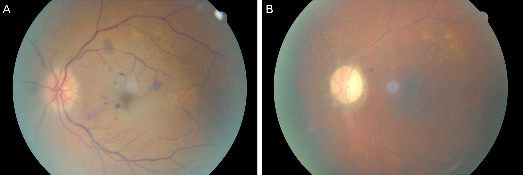
Figure 3. (A) Fundus photograph of the left eye showing retinal hemorrhages, disc edema and macular opacification with a cherry-red spot at the initial visit. (B) Six months later, fundus photograph of the left eye shows narrowing of the retinal arteries and optic disc pallor.

Figure 4. Fluorescein angiography of the left eye at the initial visit. (A) At 29 seconds after injection, filling of arterioles are still incomplete. (B) At 55 seconds after injection, filling of retinal vein is still incomplete. The left eye shows a delayed arm-to-retinal circulation and arteriovenous transit time.
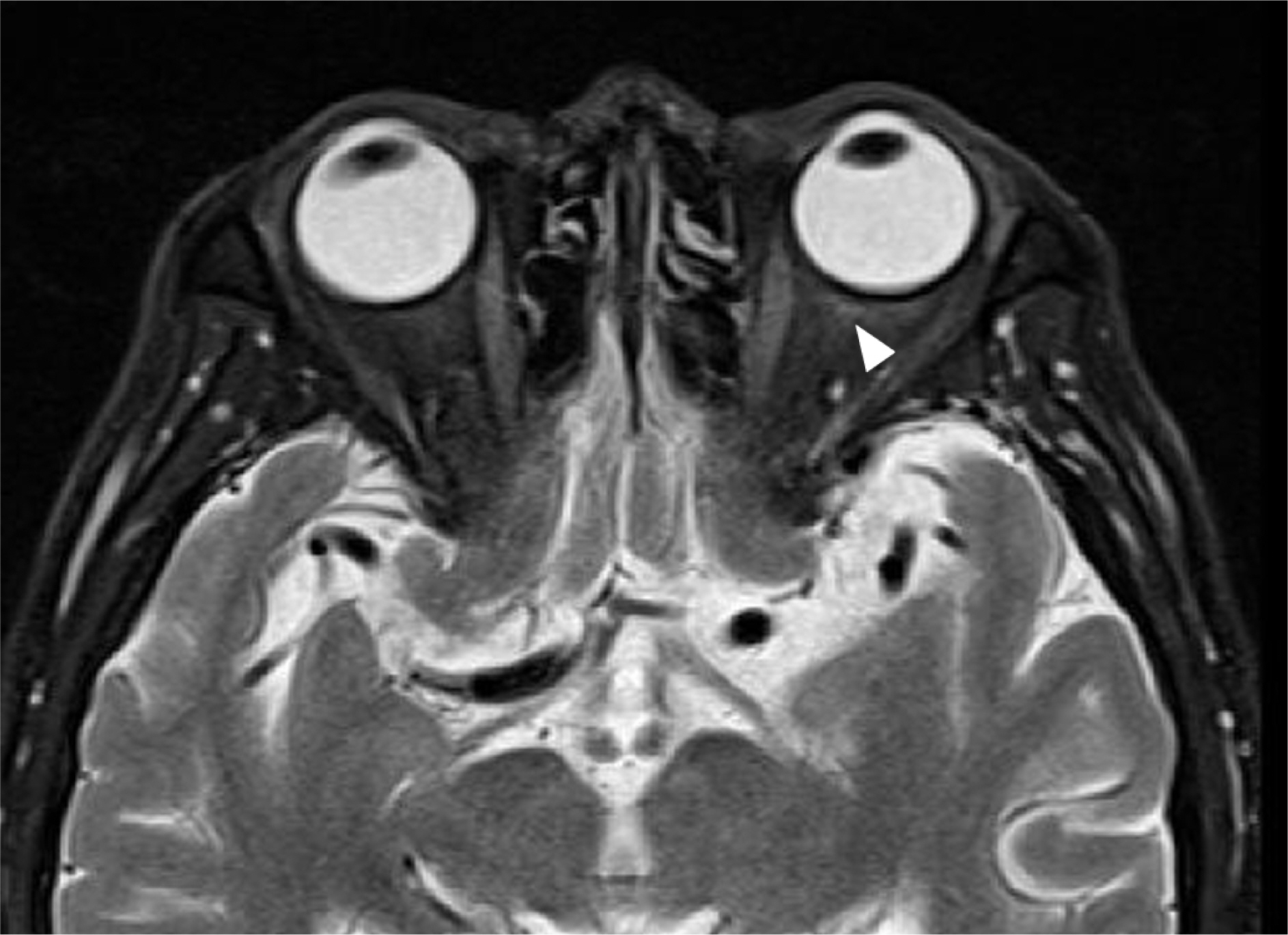
Figure 5. Magnetic resonance imaging shows slightly enhanced left optic nerve after contrast injection on T2-weighted image (white arrowhead).
Reference
-
References
1. Liesegang TJ. Herpes zoster ophthalmicus natural history, risk factors, clinical presentation, and morbidity. Ophthalmology. 2008; 115(2 Suppl):S3–12.2. Ahn M, Cho NC. Central retinal artery occlusion in herpes zoster ophthalmicus. J Pediatr Ophthalmol Strabismus. 2002; 39:123–4.
Article3. Atmaca LS, Ozmert E. Optic neuropathy and central retinal artery occlusion in a patient with herpes zoster ophthalmicus. Ann Ophthalmol. 1992; 24:50–3.4. Camuglia JE, Beltz JE, Khurana K, Hall AJ. An unusual cause of visual loss after Herpes zoster ophthalmicus: a case report. Cases J. 2010; 3:17.
Article5. Scharf Y, Kraus E, Zonis S. Optic neuropathy and central retinal artery occlusion in a case of herpes zoster ophthalmicus. Ann Ophthalmol. 1987; 19:77–8.6. Wilson CA, Wander AH, Choromokos EA. Central retinal artery obstruction in herpes zoster ophthalmicus and cerebral vasculopathy. Ann Ophthalmol. 1990; 22:347–51.7. Kim JY, Ahn M, Lee DW. Two cases of optic neuritis in herpes zoster ophthalmicus. J Korean Ophthalmol Soc. 2008; 49:1028–32.
Article8. Heininger U, Seward JF. Varicella. Lancet. 2006; 368:1365–76.
Article9. Cho GE, Choi KR, Jun RM. Herpes zoster ophthalmicus in patients younger than 50 years versus 50 years and older. J Korean Ophthalmol Soc. 2013; 54:19–25.
Article10. Kim M, Choi MY, Chae JB. Complicated ophthalmopathy in herpes zoster ophthalmicus including vitreous opacity, retinal hemorrhage and optic neuropathy. J Korean Ophthalmol Soc. 2013; 54:513–7.
Article11. Kim YR, Cho NC, You IC. Comparison of herpes zoster ophthalmicus in patients 60 years older versus younger than 60 years. J Korean Ophthalmol Soc. 2013; 54:568–73.
Article12. Han JB, Kim TG, Jin KH. Three cases of pupil abnormality in herpes zoster ophthalmicus. J Korean Ophthalmol Soc. 2013; 54:1452–7.
Article13. Elble RJ. Intracerebral hemorrhage with herpes zoster ophthalmicus. Ann Neurol. 1983; 14:591–2.
Article14. Kuroiwa Y, Furukawa T. Hemispheric infarction after herpes zoster ophthalmicus: computed tomography and angiography. Neurology. 1981; 31:1030–2.
Article15. Kim SH, Yun YJ, Kim JY. Central retinal artery occlusion associated with chickenpox. J Korean Ophthalmol Soc. 2008; 49:853–7.
Article16. Nam YH, Im M, Lee EJ, et al. A case of central retinal artery occlusion associated with chickenpox. Korean J Dermatol. 2004; 42:1337–9.
