Full mouth rehabilitation of a patient with difficulties in guiding centric relation: A case report
- Affiliations
-
- 1Department of Prosthodontics, School of Dentistry, Chonnaml National University, Gwangju, Republic of Korea. msvang@gmail.com
- KMID: 2118810
- DOI: http://doi.org/10.4047/jkap.2015.53.4.366
Abstract
- The Gothic arch tracing method using a Gothic arch tracer which is one of the centric relation recording methods can reproduce mandibular movement more accurately by describing the path of mandibular curvilinear motion. This case reports that we have satisfactory results by recording reproducible centric relation using a gothic arch tracing method in a patient who has difficulty to induce centric relation by operator due to parafunctional movement.
Figure
-
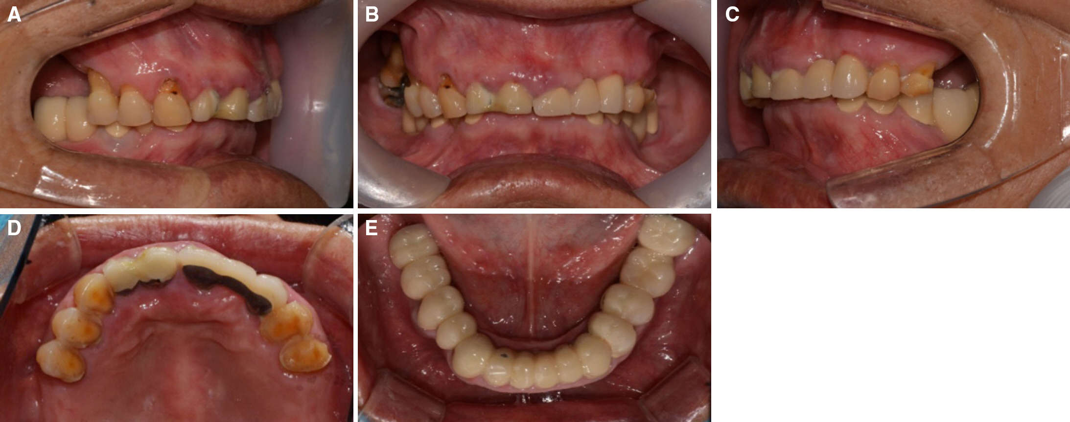
Fig. 1. Initial intraoral photographs. Severe attrition observed on occlusal surface. (A) Right side view, (B) Frontal view, (C) Left side view, (D) Maxillary occlusal view,(E) Mandibular occlusal view.

Fig. 2. Initial panoramic radiograph.
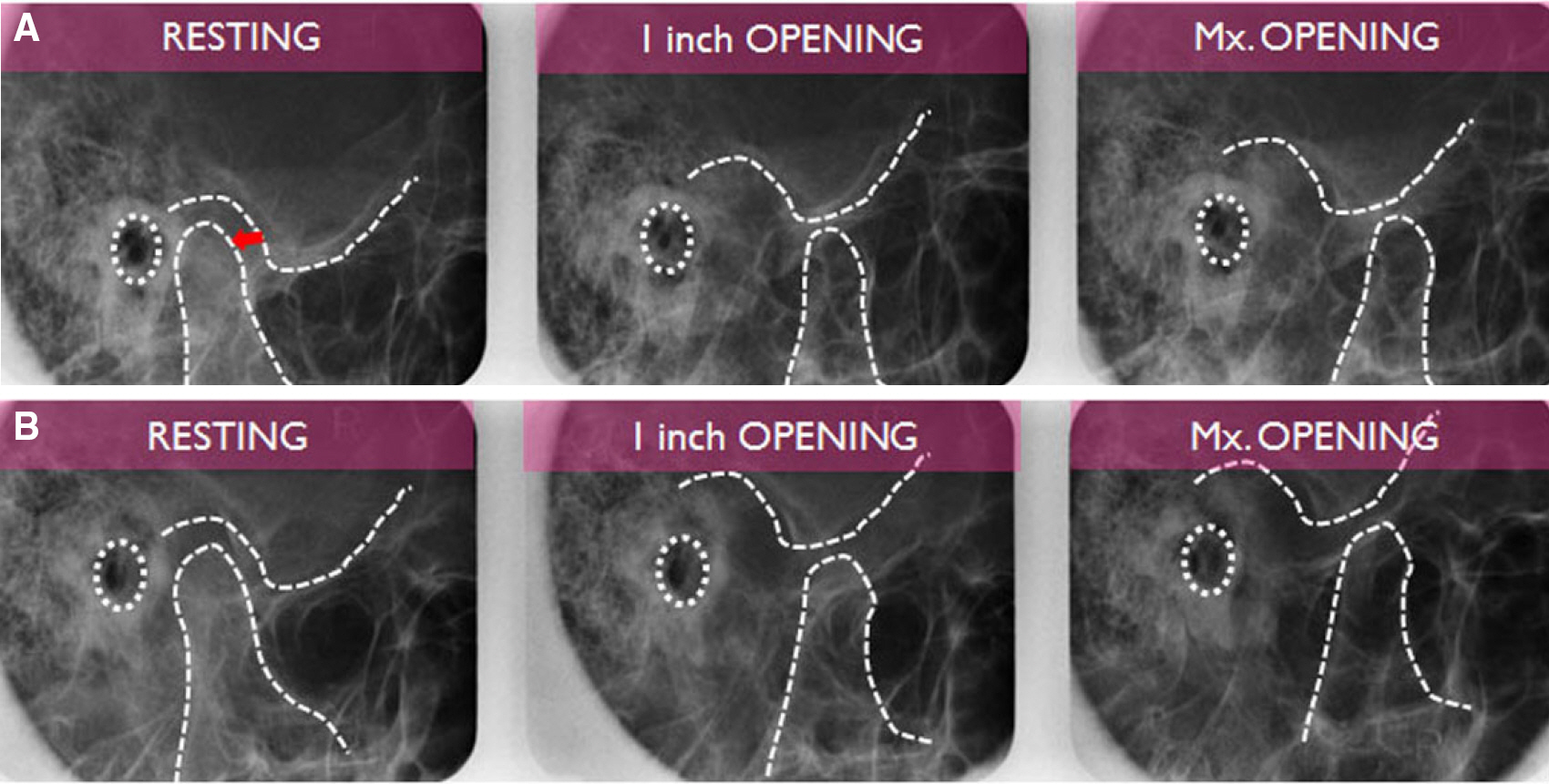
Fig. 3. Initial transcranial view of both TMJ. (A) Left TMJ. The arrow indicates retrude position of condyle. Internal derangement with reduction on the left TMJ, (B) Right TMJ.
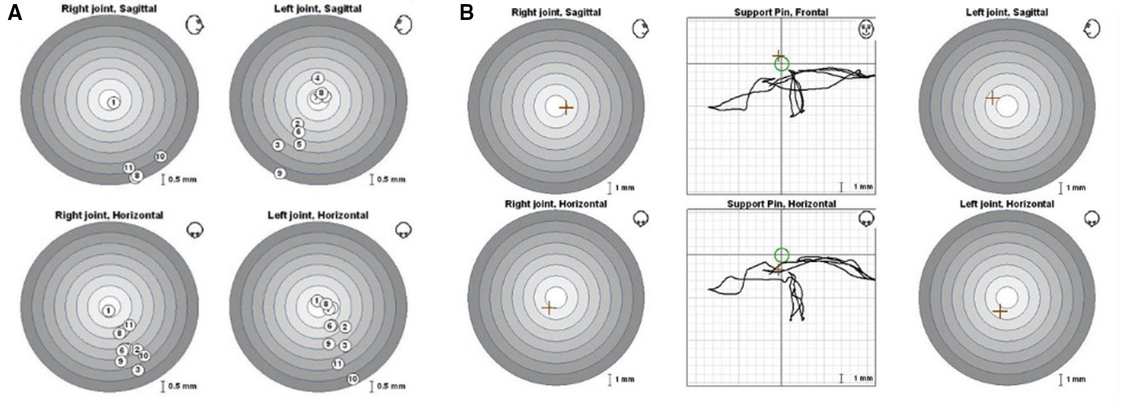
Fig. 4. Evaluation with ARCUS® digma II. (A) EPA test. ① indicates MICP. Others indicates unstable centric relation, (B) Gothic arch tracing. There is no reproducibility due to parafunctional habitual movement.
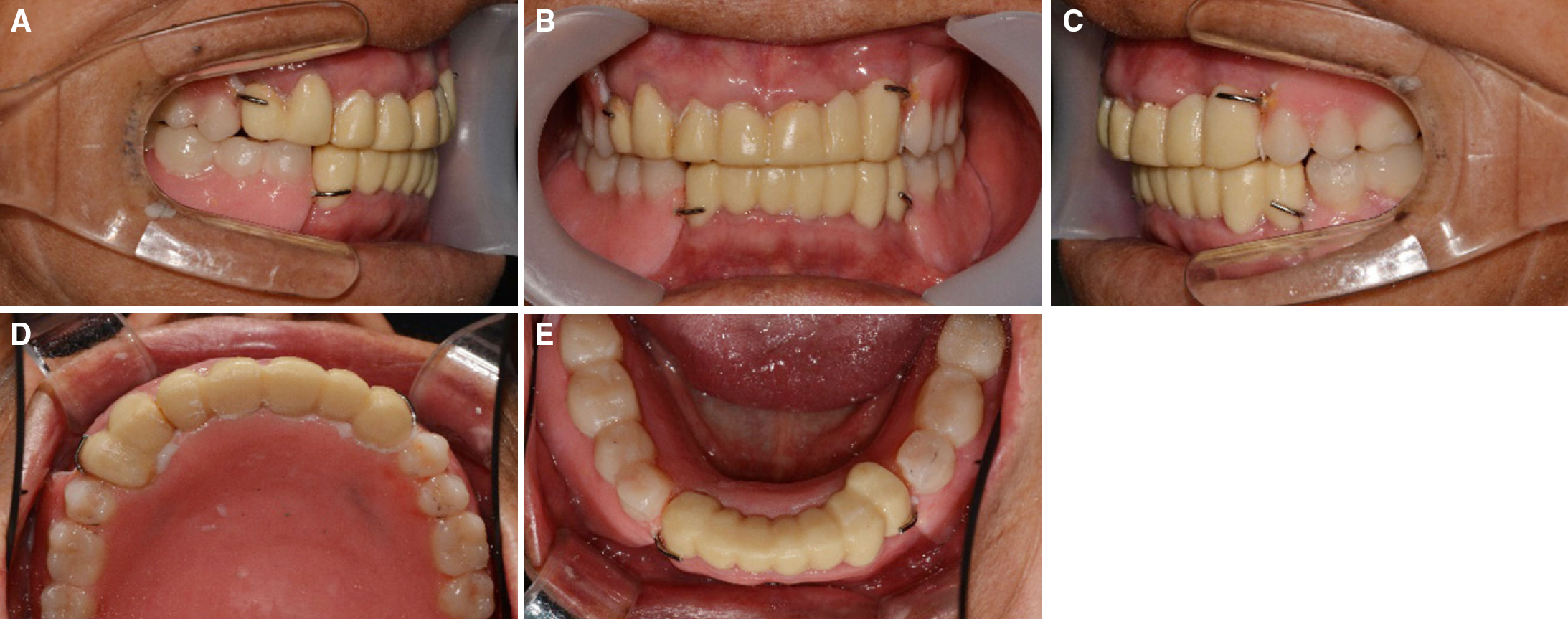
Fig. 5. Intraoral photographs after first provisional restoration placement. (A) Right side view, (B) Frontal view, (C) Left side view, (D) Maxillary occlusal view, (E) Mandibular occlusal view. Monoplane occlusion applied for treatment denture.

Fig. 6. Jaw relation taking with Gothic arch tracer. (A) Gothic arch tracer applied into patient mouth, (B) Repeat until taking stable and reproducible graph, (C) Confirm the ideal graph.
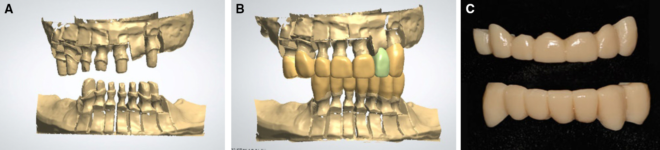
Fig. 7. CAD/CAM for second provisional restoration. (A) Model scan, (B) Wax up scan, (C) second provisional restoration.

Fig. 8. Intraoral and extraoral photographs after second provisional restoration placement. (A) Right side view, (B) Frontal view, (C) Left side view, (D) Maxillary occlusal view, (E) Mandibular occlusal view, (F) Extraoral photograph.
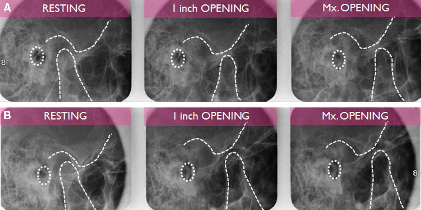
Fig. 9. Transcranial view of both TMJ after second provisional restoration placement. WNL on both TMJ. (A) Left TMJ, (B) Right TMJ.

Fig. 10. Evaluation with ARCUS® digma II. (A) EPA test. ① indicates MICP. Others indicates stable centric relation, (B) Gothic arch tracing. There is reproducibility due to stable centric relation.
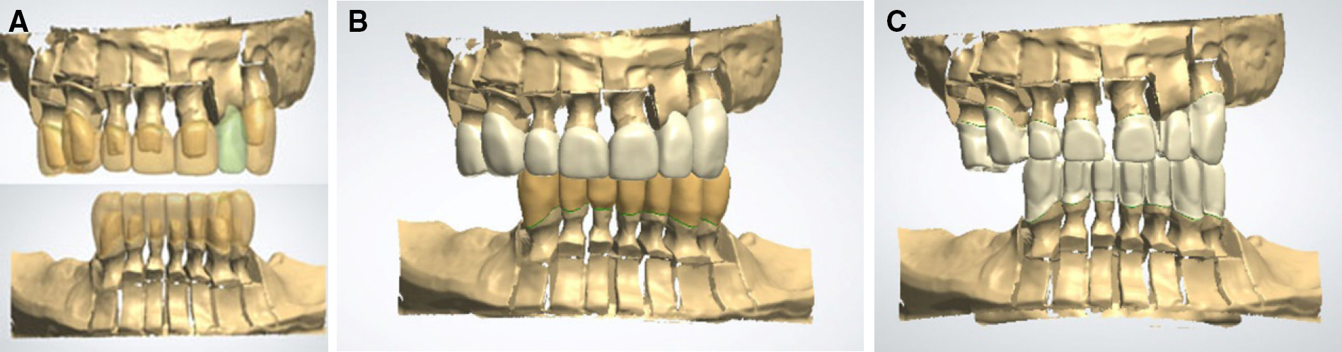
Fig. 11. CAD/CAM for surveyed PFM crowns. (A) Superimposition of wax up and die model, (B) CAD for surveyed PFM crowns, (C) Cutback on CAD for metal coping fabrication.
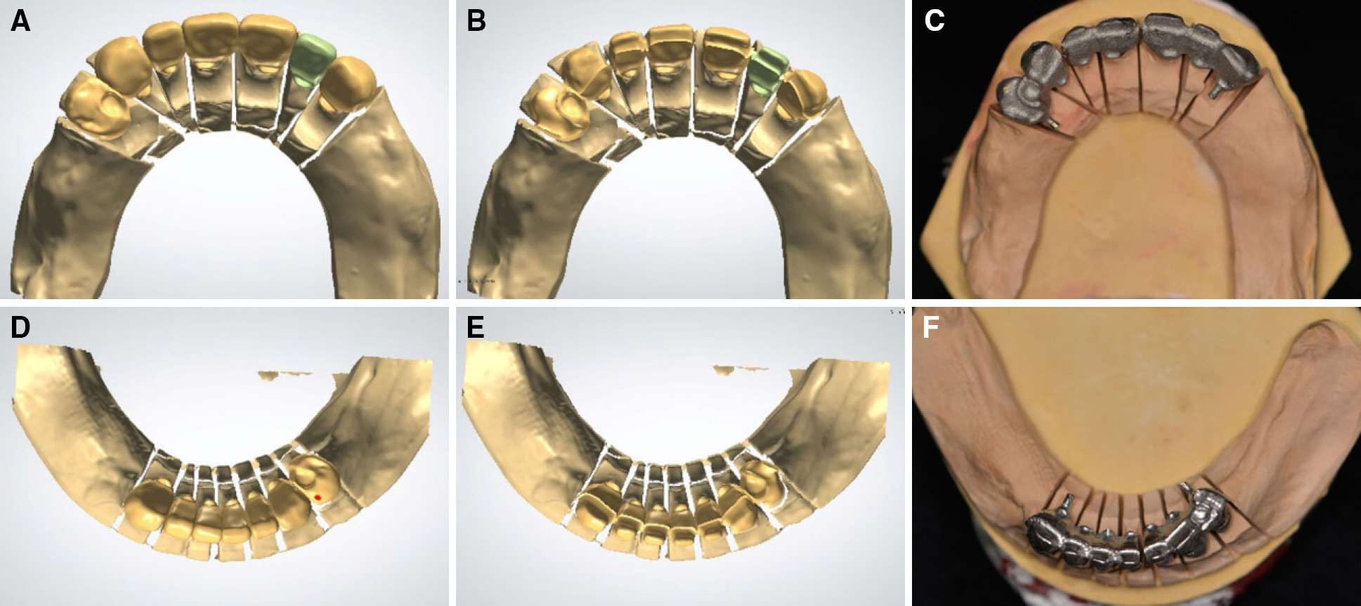
Fig. 12. CAD/CAM for metal coping of surveyed PFM crowns. (A, D) Design of surveyed PFM crowns, (B, E) 1.2 mm uniformly cutback on CAD for porcelain veneering, (C, F) metal coping fabrication with Co-Cr block.
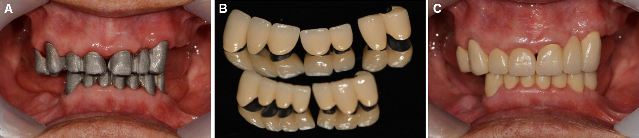
Fig. 13. Metal coping try-in and surveyed PFM crowns placement. (A) Metal coping try-in, (B) Porcelain veneering on metal coping, (C) Surveyed PFM crown placement.
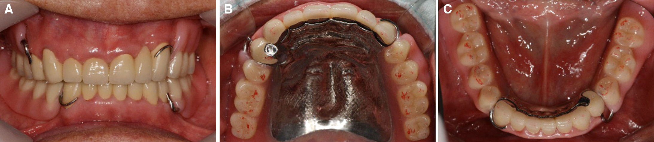
Fig. 14. Definitive denture placement and check the bite. (A) Definitive denture placement, (B) Check the bite on maxilla, (C) Check the bite on mandible.
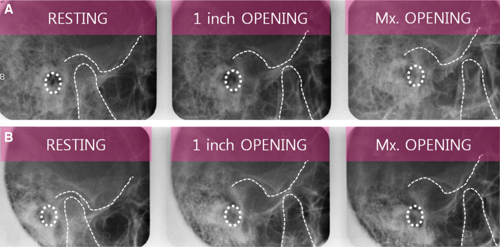
Fig. 15. Transcranial view of both TMJ after definitive denture placement. WNL on both TMJ. (A) Left TMJ, (B) Right TMJ.

Fig. 16. Evaluation with ARCUS® digma II. (A) EPA test. ① indicates MICP. Others indicates stable centric relation. MICP synchronize with centric relation, (B) Gothic arch tracing. There is reproducibility due to stable centric relation.
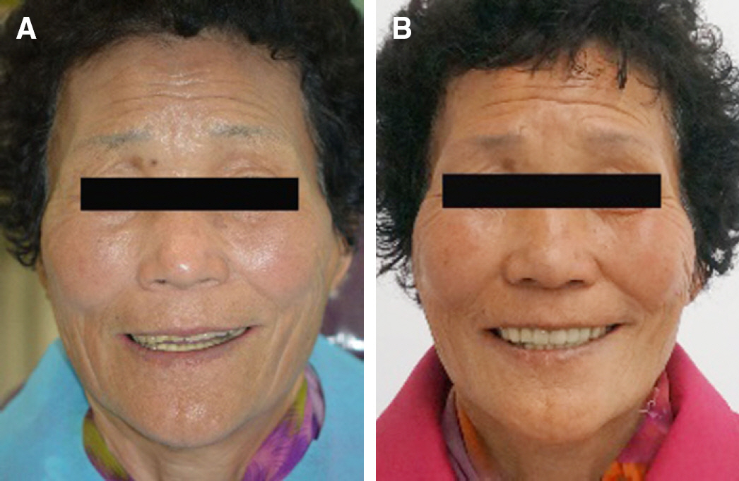
Fig. 17. Patient’ s profile. (A) Profile of first visit, (B) Profile of after definitive prosthesis placement.
Reference
-
1. The glossary of prosthodontic terms. J Prosthet Dent. 2005; 94:10–92.2. Dawson PE. New definition for relating occlusion to varying conditions of the temporomandibular joint. J Prosthet Dent. 1995; 74:619–27.
Article3. Kantor ME, Silverman SI, Garfinkel L. Centric-relation recording techniques-a comparative investigation. J Prosthet Dent. 1972; 28:593–600.
Article4. Wojdyla SM, Wiederhold DM. Using intraoral Gothic arch tracing to balance full dentures and determine centric relation and occlusal vertical dimension. Dent Today. 2005; 24:74–7.5. Abrahamsen TC. The worn dentition-pathognomonic patterns of abrasion and erosion. Int Dent J. 2005; 55:268–76.6. Schellhas KP, Piper MA, Omlie MR. Facial skeleton remodeling due to temporomandibular joint degeneration: an imaging study of 100 patients. AJNR Am J Neuroradiol. 1990; 11:541–51.
Article7. Lytle JD. Clinician’ s index of occlusal disease: definition, recognition, and management. Int J Periodontics Restorative Dent. 1990; 10:102–23.8. The Korean academy of stomatognathic function & occlusion. Textbook of human jaw function & occlusion. Yenang, Seoul. 2014. 130.9. Williamson EH, Lundquist DO. Anterior guidance: its effect on electromyographic activity of the temporal and masseter muscles. J Prosthet Dent. 1983; 49:816–23.
Article10. Dawson PE. Centric relation. Its effect on occluso-muscle har-mony. Dent Clin North Am. 1979; 23:169–80.11. Lucia VO. Principles of articulation. Dent Clin North Am. 1979; 23:199–211.12. Carroll WJ, Woelfel JB, Huffman RW. Simple application of anterior jig or leaf gauge in routine clinical practice. J Prosthet Dent. 1988; 59:611–7.
Article13. Yurkstas AA, Kapur KK. Factors influencing centric relation records in edentulous mouths. J Prosthet Dent. 2005; 93:305–10.
Article14. Gysi A, Wayne OT. The problem of articulation. Dental Cosmos. 1910; 52:403–10.15. In-Ho Cho, et al. Prosthodontic treatment for edentulous patients. 2nd ed, Yenang, Seoul, Korea. 2014. 235.16. Thakur M, Jain V, Parkash H, Kumar P. A comparative evaluation of static and functional methods for recording centric relation and condylar guidance: a clinical study. J Indian Prosthodont Soc. 2012; 12:175–81.
Article17. Myers ML. Centric relation records-historical review. J Prosthet Dent. 1982; 47:141–5.
Article18. Trapozzano VR. A comparison of the equalization of pressure by means of the central bearing point and wax check bites. J Am Dent Assoc. 1949; 38:586–91.
Article19. Payne SH. Selective occlusion. J Prosthet Dent. 1955; 73:301–4.
Article20. Pont GD. A balancing appliance for complete dentures. J Prosthet Dent. 1961; 11:434–9.
Article21. Kim YJ, et al. Biofunctional Prosthetic system. Jisung, Seoul, Korea;. 2004. 62–9.22. Jones PM. The monoplane occlusion for complete dentures. J Am Dent Assoc. 1972; 85:94–100.
Article
- Full Text Links
-
- Actions
-
Cited
- CITED
-
- Close
- Share
-
- Similar articles
-
- A case of full mouth rehabilitation with orthodontic treatment in patient with extensive tooth erosion and wear using monolithic zirconia prostheses
- Full mouth rehabilitation of a patient with tooth wear and insufficient restorative space due to loss of posterior teeth support: a case report
- Prosthetic Rehabilitation in Patients with Severe Hearing Disability and Loss of Occlusal Vertical Dimension: A Case Report
- Full mouth rehabilitation using removable prosthesis of patient with unstable mandible movements: A case report
- Full mouth rehabilitation in patient with loss of vertical dimension and deep bite due to tooth wear
