J Adv Prosthodont.
2014 Jun;6(3):151-156. 10.4047/jap.2014.6.3.151.
Tensile strength of bilayered ceramics and corresponding glass veneers
- Affiliations
-
- 1Department of Prosthodontics, Faculty of Dentistry, Mahidol University, Bangkok, Thailand. Chuchai.anu@mahidol.edu
- 2Department of Advanced General Dentistry, Faculty of Dentistry, Mahidol University, Bangkok, Thailand.
- KMID: 2118224
- DOI: http://doi.org/10.4047/jap.2014.6.3.151
Abstract
- PURPOSE
To investigate the microtensile bond strength between two all-ceramic systems; lithium disilicate glass ceramic and zirconia core ceramics bonded with their corresponding glass veneers.
MATERIALS AND METHODS
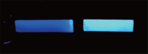
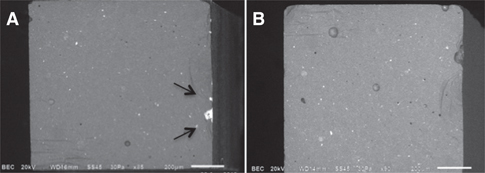
Blocks of core ceramics (IPS e.max(R) Press and Lava(TM) Frame) were fabricated and veneered with their corresponding glass veneers. The bilayered blocks were cut into microbars; 8 mm in length and 1 mm2 in cross-sectional area (n = 30/group). Additionally, monolithic microbars of these two veneers (IPS e.max(R) Ceram and Lava(TM) Ceram; n = 30/group) were also prepared. The obtained microbars were tested in tension until fracture, and the fracture surfaces of the microbars were examined with fluorescent black light and scanning electron microscope (SEM) to identify the mode of failure. One-way ANOVA and the Dunnett's T3 test were performed to determine significant differences of the mean microtensile bond strength at a significance level of 0.05.
RESULTS
The mean microtensile bond strength of IPS e.max(R) Press/IPS e.max(R) Ceram (43.40 +/- 5.51 MPa) was significantly greater than that of Lava(TM) Frame/Lava(TM) Ceram (31.71 +/- 7.03 MPa)(P<.001). Fluorescent black light and SEM analysis showed that most of the tested microbars failed cohesively in the veneer layer. Furthermore, the bond strength of Lava(TM) Frame/Lava(TM) Ceram was comparable to the tensile strength of monolithic glass veneer of Lava(TM) Ceram, while the bond strength of bilayered IPS e.max(R) Press/IPS e.max(R) Ceram was significantly greater than tensile strength of monolithic IPS e.max(R) Ceram.
CONCLUSION
Because fracture site occurred mostly in the glass veneer and most failures were away from the interfacial zone, microtensile bond test may not be a suitable test for bonding integrity. Fracture mechanics approach such as fracture toughness of the interface may be more appropriate to represent the bonding quality between two materials.
Keyword
Figure
Reference
-
1. Raigrodski AJ. Contemporary materials and technologies for all-ceramic fixed partial dentures: a review of the literature. J Prosthet Dent. 2004; 92:557–562.2. Conrad HJ, Seong WJ, Pesun IJ. Current ceramic materials and systems with clinical recommendations: a systematic review. J Prosthet Dent. 2007; 98:389–404.3. Odman P, Andersson B. Procera AllCeram crowns followed for 5 to 10.5 years: a prospective clinical study. Int J Prosthodont. 2001; 14:504–509.4. Raigrodski AJ, Chiche GJ, Potiket N, Hochstedler JL, Mohamed SE, Billiot S, Mercante DE. The efficacy of posterior three-unit zirconium-oxide-based ceramic fixed partial dental prostheses: a prospective clinical pilot study. J Prosthet Dent. 2006; 96:237–244.5. Sailer I, Pjetursson BE, Zwahlen M, Hämmerle CH. A systematic review of the survival and complication rates of all-ceramic and metal-ceramic reconstructions after an observation period of at least 3 years. Part II: Fixed dental prostheses. Clin Oral Implants Res. 2007; 18:86–96.6. Vult von Steyern P, Carlson P, Nilner K. All-ceramic fixed partial dentures designed according to the DC-Zirkon technique. A 2-year clinical study. J Oral Rehabil. 2005; 32:180–187.7. Guazzato M, Proos K, Sara G, Swain MV. Strength, reliability, and mode of fracture of bilayered porcelain/core ceramics. Int J Prosthodont. 2004; 17:142–149.8. Dündar M, Ozcan M, Gökçe B, Cömlekoğlu E, Leite F, Valandro LF. Comparison of two bond strength testing methodologies for bilayered all-ceramics. Dent Mater. 2007; 23:630–636.9. Della Bona A, van Noort R. Shear vs. tensile bond strength of resin composite bonded to ceramic. J Dent Res. 1995; 74:1591–1596.10. El Zohairy AA, de Gee AJ, de Jager N, van Ruijven LJ, Feilzer AJ. The influence of specimen attachment and dimension on microtensile strength. J Dent Res. 2004; 83:420–424.11. Sano H, Shono T, Sonoda H, Takatsu T, Ciucchi B, Carvalho R, Pashley DH. Relationship between surface area for adhesion and tensile bond strength-evaluation of a micro-tensile bond test. Dent Mater. 1994; 10:236–240.12. Aboushelib MN, Kleverlaan CJ, Feilzer AJ. Effect of zirconia type on its bond strength with different veneer ceramics. J Prosthodont. 2008; 17:401–408.13. El Zohairy AA, Saber MH, Abdalla AI, Feilzer AJ. Efficacy of microtensile versus microshear bond testing for evaluation of bond strength of dental adhesive systems to enamel. Dent Mater. 2010; 26:848–854.14. Ferrari M, Goracci C, Sadek F, Eduardo P, Cardoso C. Microtensile bond strength tests: scanning electron microscopy evaluation of sample integrity before testing. Eur J Oral Sci. 2002; 110:385–391.15. Aboushelib MN, de Jager N, Kleverlaan CJ, Feilzer AJ. Microtensile bond strength of different components of core veneered all-ceramic restorations. Dent Mater. 2005; 21:984–991.16. Aboushelib MN, de Kler M, van der Zel JM, Feilzer AJ. Microtensile bond strength and impact energy of fracture of CAD-veneered zirconia restorations. J Prosthodont. 2009; 18:211–216.17. Aboushelib MN, Kleverlaan CJ, Feilzer AJ. Microtensile bond strength of different components of core veneered all-ceramic restorations. Part II: Zirconia veneering ceramics. Dent Mater. 2006; 22:857–863.18. Taskonak B, Borges GA, Mecholsky JJ Jr, Anusavice KJ, Moore BK, Yan J. The effects of viscoelastic parameters on residual stress development in a zirconia/glass bilayer dental ceramic. Dent Mater. 2008; 24:1149–1155.19. Taskonak B, Mecholsky JJ Jr, Anusavice KJ. Residual stresses in bilayer dental ceramics. Biomaterials. 2005; 26:3235–3241.20. Tan JP, Sederstrom D, Polansky JR, McLaren EA, White SN. The use of slow heating and slow cooling regimens to strengthen porcelain fused to zirconia. J Prosthet Dent. 2012; 107:163–169.21. Aboushelib MN, Kleverlaan CJ, Feilzer AJ. Microtensile bond strength of different components of core veneered all-ceramic restorations. Part 3: double veneer technique. J Prosthodont. 2008; 17:9–13.22. Benetti P, Della Bona A, Kelly JR. Evaluation of thermal compatibility between core and veneer dental ceramics using shear bond strength test and contact angle measurement. Dent Mater. 2010; 26:743–750.23. Benetti P, Pelogia F, Valandro LF, Bottino MA, Bona AD. The effect of porcelain thickness and surface liner application on the fracture behavior of a ceramic system. Dent Mater. 2011; 27:948–953.24. Scherrer SS, Cesar PF, Swain MV. Direct comparison of the bond strength results of the different test methods: a critical literature review. Dent Mater. 2010; 26:e78–e93.25. Tam LE, Pilliar RM. Fracture toughness of dentin/resin-composite adhesive interfaces. J Dent Res. 1993; 72:953–959.26. Ruse ND, Troczynski T, MacEntee MI, Feduik D. Novel fracture toughness test using a notchless triangular prism (NTP) specimen. J Biomed Mater Res. 1996; 31:457–463.27. Tam LE, Khoshand S, Pilliar RM. Fracture resistance of dentin-composite interfaces using different adhesive resin layers. J Dent. 2001; 29:217–225.28. Anunmana C, Anusavice KJ, Mecholsky JJ Jr. Interfacial toughness of bilayer dental ceramics based on a short-bar, chevron-notch test. Dent Mater. 2010; 26:111–117.29. Pashley DH, Sano H, Ciucchi B, Yoshiyama M, Carvalho RM. Adhesion testing of dentin bonding agents: a review. Dent Mater. 1995; 11:117–125.30. Phrukkanon S, Burrow MF, Tyas MJ. The influence of cross-sectional shape and surface area on the microtensile bond test. Dent Mater. 1998; 14:212–221.
- Full Text Links
- Actions
-
Cited
- CITED
-
- Close
- Share
- Similar articles
-
- Tensile bond strength of glass ionomer cements
- Biaxial flexural strength of bilayered zirconia using various veneering ceramics
- Mechanical Properties Of Reused Lithium Disilicate Glass-Ceramic Of Ips Empress 2 System
- The effects of salivary contamination on tensile bond strength of resin modified glass ionomer cements in bonding brackets
- EFFECT OF CeCO2 ADDITION IN GLASS COMPOSITION ON THE STRENGTH OF ALUMINA-GLASS COMPOSITES


