Imaging Sci Dent.
2015 Mar;45(1):41-47. 10.5624/isd.2015.45.1.41.
Accuracy and reliability of stitched cone-beam computed tomography images
- Affiliations
-
- 1Private Practice, Reconstructive Dental Specialists of Utah, Salt Lake City, UT, USA.
- 2Department of Prosthodontics, University of Tennessee Health Science Center College of Dentistry, Memphis, TN, USA. sahuja@uthsc.edu
- KMID: 2116790
- DOI: http://doi.org/10.5624/isd.2015.45.1.41
Abstract
- PURPOSE
This study was performed to evaluate the linear distance accuracy and reliability of stitched small field of view (FOV) cone-beam computed tomography (CBCT) reconstructed images for the fabrication of implant surgical guides.
MATERIALS AND METHODS
Three gutta percha points were fixed on the inferior border of a cadaveric mandible to serve as control reference points. Ten additional gutta percha points, representing fiduciary markers, were scattered on the buccal and lingual cortices at the level of the proposed complete denture flange. A digital caliper was used to measure the distance between the reference points and fiduciary markers, which represented the anatomic linear dimension. The mandible was scanned using small FOV CBCT, and the images were then reconstructed and stitched using the manufacturer's imaging software. The same measurements were then taken with the CBCT software.
RESULTS
The anatomic linear dimension measurements and stitched small FOV CBCT measurements were statistically evaluated for linear accuracy. The mean difference between the anatomic linear dimension measurements and the stitched small FOV CBCT measurements was found to be 0.34 mm with a 95% confidence interval of +0.24 - +0.44 mm and a mean standard deviation of 0.30 mm. The difference between the control and the stitched small FOV CBCT measurements was insignificant within the parameters defined by this study.
CONCLUSION
The proven accuracy of stitched small FOV CBCT data sets may allow image-guided fabrication of implant surgical stents from such data sets.
MeSH Terms
Figure
-

Fig. 1 Control measurement is performed using digital calipers.
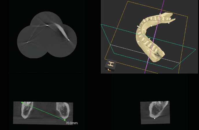
Fig. 2 A Stitched image using the small field of view cone-beam computed tomography images is seen. The measurement is performed using the multiplanar view after locating the reference points and markers accurately.
Cited by 1 articles
-
The accuracy evaluation of digital surgical stents according to supported type
Junyoun Lee, Minho Yoon, Taeseok Park, Inkon Chun, Kwidug Yun
J Korean Acad Prosthodont. 2018;56(1):8-16. doi: 10.4047/jkap.2018.56.1.8.
Reference
-
1. BouSerhal C, Jacobs R, Quirynen M, van Steenberghe D. Imaging technique selection for the preoperative planning of oral implants: a review of the literature. Clin Implant Dent Relat Res. 2002; 4:156–172.
Article2. Aranyarachkul P, Caruso J, Gantes B, Schulz E, Riggs M, Dus I, et al. Bone density assessments of dental implant sites: 2. Quantitative cone-beam computerized tomography. Int J Oral Maxillofac Implants. 2005; 20:416–424.3. Greenstein G, Tarnow D. The mental foramen and nerve: clinical and anatomical factors related to dental implant placement: a literature review. J Periodontol. 2006; 77:1933–1943.
Article4. Lee S, Gantes B, Riggs M, Crigger M. Bone density assessments of dental implant sites: 3. Bone quality evaluation during osteotomy and implant placement. Int J Oral Maxillofac Implants. 2007; 22:208–212.5. Kraut RA. Interactive CT diagnostics, planning and preparation for dental implants. Implant Dent. 1998; 7:19–25.
Article6. Sarment DP, Sukovic P, Clinthorne N. Accuracy of implant placement with a stereolithographic surgical guide. Int J Oral Maxillofac Implants. 2003; 18:571–577.7. Parel SM, Triplett RG. Interactive imaging for implant planning, placement, and prosthesis construction. J Oral Maxillofac Surg. 2004; 62:41–47.
Article8. Guerrero ME, Jacobs R, Loubele M, Schutyser F, Suetens P, van Steenberghe D. State-of-the-art on cone beam CT imaging for preoperative planning of implant placement. Clin Oral Investig. 2006; 10:1–7.
Article9. Balshi SF, Wolfinger GJ, Balshi TJ. Surgical planning and prosthesis construction using computer technology and medical imaging for immediate loading of implants in the pterygomaxillary region. Int J Periodontics Restorative Dent. 2006; 26:239–247.10. Nikzad S, Azari A. A novel stereolithographic surgical guide template for planning treatment involving a mandibular dental implant. J Oral Maxillofac Surg. 2008; 66:1446–1454.
Article11. Valente F, Schiroli G, Sbrenna A. Accuracy of computer-aided oral implant surgery: a clinical and radiographic study. Int J Oral Maxillofac Implants. 2009; 24:234–242.12. Jung RE, Schneider D, Ganeles J, Wismeijer D, Zwahlen M, Hämmerle CH, et al. Computer technology applications in surgical implant dentistry: a systematic review. Int J Oral Maxillofac Implants. 2009; 24:92–109.
Article13. Sanna A, Molly ML, van Steenberghe D. Immediately loaded CAD-CAM manufactured fixed complete dentures using flapless implant placement procedures: a cohort study of consecutive patients. J Prosthet Dent. 2007; 97:331–339.
Article14. Balshi TJ, Wolfinger GJ. Teeth in a day for the maxilla and mandible: case report. Clin Implant Dent Relat Res. 2003; 5:11–16.
Article15. Hultin M, Svensson K, Trulsson M. Clinical advantages of computer-guided implant placement: a systematic review. Clin Oral Implants Res. 2012; 23:124–135.
Article16. Ganz SD. Computer-aided design/computer-aided manufacturing applications using CT and cone beam CT scanning technology. Dent Clin North Am. 2008; 52:777–808.
Article17. Lindh C, Petersson A, Klinge B. Measurements of distances related to the mandibular canal in radiographs. Clin Oral Implants Res. 1995; 6:96–103.
Article18. Kobayashi K, Shimoda S, Nakagawa Y, Yamamoto A. Accuracy in measurement of distance using limited cone-beam computerized tomography. Int J Oral Maxillofac Implants. 2004; 19:228–231.19. Kopp S, Ottl P. Dimensional stability in composite cone beam computed tomography. Dentomaxillofac Radiol. 2010; 39:512–516.
Article20. Berco M, Rigali PH Jr, Miner RM, DeLuca S, Anderson NK, Will LA. Accuracy and reliability of linear cephalometric measurements from cone-beam computed tomography scans of a dry human skull. Am J Orthod Dentofacial Orthop. 2009; 136:17e1–e9.
Article21. Hassan B, van der Stelt P, Sanderink G. Accuracy of three-dimensional measurements obtained from cone beam computed tomography surface-rendered images for cephalometric analysis: influence of patient scanning position. Eur J Orthod. 2009; 31:129–134.
Article22. Lascala CA, Panella J, Marques MM. Analysis of the accuracy of linear measurements obtained by cone beam computed tomography (CBCT-NewTom). Dentomaxillofac Radiol. 2004; 33:291–294.
Article23. Periago DR, Scarfe WC, Moshiri M, Scheetz JP, Silveira AM, Farman AG. Linear accuracy and reliability of cone beam CT derived 3-dimensional images constructed using an orthodontic volumetric rendering program. Angle Orthod. 2008; 78:387–395.
Article24. Scarfe WC, Li Z, Aboelmaaty W, Scott SA, Farman AG. Maxillofacial cone beam computed tomography: essence, elements and steps to interpretation. Aust Dent J. 2012; 57:46–60.
Article25. Mayo JR. High resolution computed tomography. Technical aspects. Radiol Clin North Am. 1991; 29:1043–1049.
- Full Text Links
-
- Actions
-
Cited
- CITED
-
- Close
- Share
-
- Similar articles
-
- Diagnostic performance of stitched and non-stitched cross-sectional cone-beam computed tomography images of a non-displaced fracture of ovine mandibular bone
- The accuracy of the imaging reformation of cone beam computed tomography for the assessment of bone defect healing
- Expression of beam hardening artifacts on horizontally stitched cone-beam computed tomography images
- Effect of Voxel Size on the Accuracy of Landmark Identification in Cone-Beam Computed Tomography Images
- Inter-observer reliability in cone-beam computed tomography assessment of the retromolar canal: A practical plan to improve diagnostic imaging
