Volumetric quantification of bone-implant contact using micro-computed tomography analysis based on region-based segmentation
- Affiliations
-
- 1Interdisciplinary Program in Radiation, Applied Life Science Major, College of Medicine, BK21, and Dental Research Institute, Seoul National University, Korea.
- 2Department of Oral and Maxillofacial Radiology, BK21, and Dental Research Institute, School of Dentistry, Seoul National University, Korea. wjyi@snu.ac.kr
- 3Department of Periodontology and Dental Research Institute, School of Dentistry, Seoul National University, Korea. periopf@snu.ac.kr
- KMID: 2116786
- DOI: http://doi.org/10.5624/isd.2015.45.1.7
Abstract
- PURPOSE
We have developed a new method of segmenting the areas of absorbable implants and bone using region-based segmentation of micro-computed tomography (micro-CT) images, which allowed us to quantify volumetric bone-implant contact (VBIC) and volumetric absorption (VA).
MATERIALS AND METHODS
The simple threshold technique generally used in micro-CT analysis cannot be used to segment the areas of absorbable implants and bone. Instead, a region-based segmentation method, a region-labeling method, and subsequent morphological operations were successively applied to micro-CT images. The three-dimensional VBIC and VA of the absorbable implant were then calculated over the entire volume of the implant. Two-dimensional (2D) bone-implant contact (BIC) and bone area (BA) were also measured based on the conventional histomorphometric method.
RESULTS
VA and VBIC increased significantly with as the healing period increased (p<0.05). VBIC values were significantly correlated with VA values (p<0.05) and with 2D BIC values (p<0.05).
CONCLUSION
It is possible to quantify VBIC and VA for absorbable implants using micro-CT analysis using a region-based segmentation method.
Keyword
MeSH Terms
Figure
-
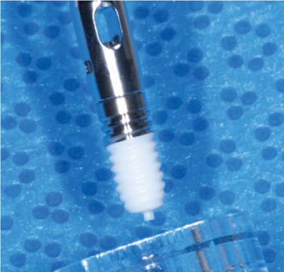
Fig. 1 An absorbable implant consists of a titanium upper part and a polylactic acid/tricalcium phosphate (PLA-TCP) lower part.
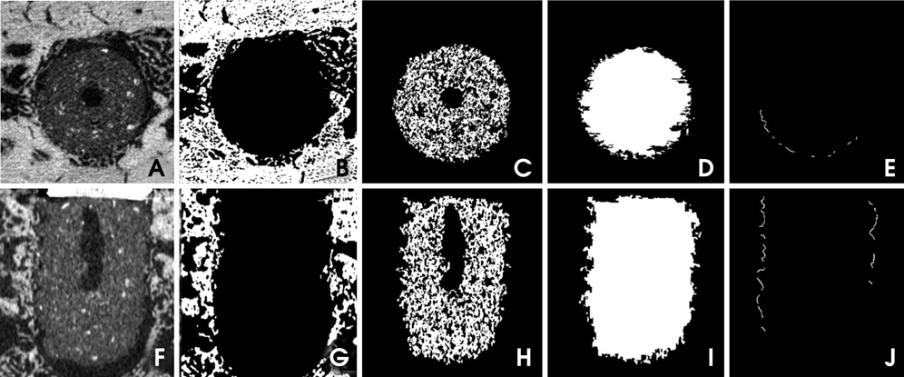
Fig. 2 Smoothed micro-computed tomography image of the absorbable implant in axial (A) and coronal (F) slices. Segmented images of the bone in axial (B) and coronal (G) slices. Segmented images of the implant in axial (C) and coronal (H) slices. Hole-filled images of the implant in axial (D) and coronal (I) slices. The implant surface in direct contact with the bone in axial (E) and coronal (J) slices

Fig. 3 Three-dimensional rendering of the reconstructed volumes shows the bone and implant after segmentation.

Fig. 4 Histomorphometric images show the absorbable implant (100×) at four weeks (A), eight weeks (B), and 12 weeks (C) after placement.

Fig. 5 The relationship between the volumetric bone-implant contact ratio (VBIC, %) and volumetric absorption (VA, %) (Pearson's correlation=0.46, p<0.05).
Reference
-
1. Schnitman PA, Wöhrle PS, Rubenstein JE, DaSilva JD, Wang NH. Ten-year results for Brånemark implants immediately loaded with fixed prostheses at implant placement. Int J Oral Maxillofac Implants. 1997; 12:495–503.2. Rossi E, Andreasen JO. Maxillary bone growth and implant positioning in a young patient: a case report. Int J Periodontics Restorative Dent. 2003; 23:113–119.3. Tarlow JL. The effect of adult growth on an anterior maxillary single-tooth implant: a clinical report. J Prosthet Dent. 2004; 92:213–215.
Article4. Laffargue P, Fialdes P, Frayssinet P, Rtaimate M, Hildebrand HF, Marchandise X. Adsorption and release of insulin-like growth factor-I on porous tricalcium phosphate implant. J Biomed Mater Res. 2000; 49:415–421.
Article5. Robert P, Mauduit J, Frank RM, Vert M. Biocompatibility and resorbability of a polylactic acid membrane for periodontal guided tissue regeneration. Biomaterials. 1993; 14:353–358.
Article6. Kulkarni RK, Pani KC, Neuman C, Leonard F. Polylactic acid for surgical implants. Arch Surg. 1966; 93:839–843.
Article7. Karabuda C, Ozdemir O, Tosun T, Anil A, Olgaç V. Histological and clinical evaluation of 3 different grafting materials for sinus lifting procedure based on 8 cases. J Periodontol. 2001; 72:1436–1442.
Article8. Brånemark PI, Hansson BO, Adell R, Breine U, Lindström J, Hallén O, et al. Osseointegrated implants in the treatment of the edentulous jaw. Experience from a 10-year period. Scand J Plast Reconstr Surg Suppl. 1977; 16:1–132.9. Albrektsson T, Brånemark PI, Hansson HA, Lindström J. Osseointegrated titanium implants. Requirements for ensuring a long-lasting, direct bone-to-implant anchorage in man. Acta Orthop Scand. 1981; 52:155–170.10. Kim DS, Kim DG, Park CJ, Cho LR. Histomorphometry and stability analysis of early loaded implants with two different surface conditions in beagle dogs. J Adv Prosthodont. 2009; 1:10–18.
Article11. Nkenke E, Hahn M, Weinzierl K, Radespiel-Troger M, Neukam FW, Engelke K. Implant stability and histomorphometry: a correlation study in human cadavers using stepped cylinder implants. Clin Oral Implants Res. 2003; 14:601–609.
Article12. Deguchi T, Nasu M, Murakami K, Yabuuchi T, Kamioka H, Takano-Yamamoto T. Quantitative evaluation of cortical bone thickness with computed tomographic scanning for orthodontic implants. Am J Orthod Dentofacial Orthop. 2006; 129:721.
Article13. Garetto LP, Chen J, Parr JA, Roberts WE. Remodeling dynamics of bone supporting rigidly fixed titanium implants: a histomorphometric comparison in four species including humans. Implant Dent. 1995; 4:235–243.
Article14. Roberts WE. Bone tissue interface. J Dent Educ. 1988; 52:804–809.
Article15. Le Guehennec L, Goyenvalle E, Lopez-Heredia MA, Weiss P, Amouriq Y, Layrolle P. Histomorphometric analysis of the osseointegration of four different implant surfaces in the femoral epiphyses of rabbits. Clin Oral Implants Res. 2008; 19:1103–1110.
Article16. Rebaudi A, Koller B, Laib A, Trisi P. Microcomputed tomographic analysis of the peri-implant bone. Int J Periodontics Restorative Dent. 2004; 24:316–325.17. Park HS, Kwon OW, Sung JH. Microscrew implant anchorage sliding mechanics. World J Orthod. 2005; 6:265–274.18. Schicho K, Kastner J, Klingesberger R, Seemann R, Enislidis G, Undt G, et al. Surface area analysis of dental implants using micro-computed tomography. Clin Oral Implants Res. 2007; 18:459–464.
Article19. Van Oossterwyck H, Duyck J, Vander Sloten J, Van der Perre G, Jansen J, Wevers M, et al. Use of microfocus computerized tomography as a new technique for characterizing bone tissue around oral implants. J Oral Implantol. 2000; 26:5–12.20. Bernhardt R, Kuhlisch E, Schulz MC, Eckelt U, Stadlinger B. Comparison of bone-implant contact and bone-implant volume between 2D-histological sections and 3D-SRrµCT slices. Eur Cell Mater. 2012; 23:237–248.21. Park YS, Yi KY, Lee IS, Jung YC. Correlation between microtomography and histomorphometry for assessment of implant osseointegration. Clin Oral Implants Res. 2005; 16:156–160.
Article22. Liu S, Broucek J, Virdi AS, Sumner DR. Limitations of using micro-computed tomography to predict bone-implant contact and mechanical fixation. J Microsc. 2012; 245:34–42.
Article23. Debats OA, Litjens GJ, Barentsz JO, Karssemeijer N, Huisman HJ. Automated 3-dimensional segmentation of pelvic lymph nodes in magnetic resonance images. Med Phys. 2011; 38:6178–6187.
Article24. Singh UP, Saxena K, Jain S. Semi-supervised method of multiple object segmentation with a region labeling and flood fill. Signal Image Process. 2011; 2:175–193.25. Park JW, An CH, Jeong SH, Suh JY. Osseointegration of commercial microstructured titanium implants incorporating magnesium: a histomorphometric study in rabbit cancellous bone. Clin Oral Implants Res. 2012; 23:294–300.
Article26. Piattelli M, Scarano A, Paolantonio M, Iezzi G, Petrone G, Piattelli A. Bone response to machined and resorbable blast material titanium implants: an experimental study in rabbits. J Oral Implantol. 2002; 28:2–8.
Article27. Buser D, Schenk RK, Steinemann S, Fiorellini JP, Fox CH, Stich H. Influence of surface characteristics on bone integration of titanium implants. A histomorphometric study in miniature pigs. J Biomed Mater Res. 1991; 25:889–902.
Article28. Jeong R, Marin C, Granato R, Suzuki M, Gil JN, Granjeiro JM, et al. Early bone healing around implant surfaces treated with variations in the resorbable blasting media method. A study in rabbits. Med Oral Patol Oral Cir Bucal. 2010; 15:e119–e125.
Article29. Rohner D, Tay A, Chung SM, Hutmacher DW. Interface of unloaded titanium implants in the iliac crest, fibula, and scapula: a histomorphometric and biomechanical study in the pig. Int J Oral Maxillofac Implants. 2004; 19:52–58.30. Butz F, Ogawa T, Chang TL, Nishimura I. Three-dimensional bone-implant integration profiling using micro-computed tomography. Int J Oral Maxillofac Implants. 2006; 21:687–695.31. Ko CY, Lim DH, Choi BH, Li J, Kim HS. Suggestion of new methodology for evaluation of osseointegration between implant and bone based on µ-CT images. Int J Precis Eng Man. 2010; 11:785–790.
Article
- Full Text Links
-
- Actions
-
Cited
- CITED
-
- Close
- Share
-
- Similar articles
-
- Micro-computed tomography analysis of changes in the periodontal ligament and alveolar bone proper induced by occlusal hypofunction of rat molars
- Torque and mechanical failure of orthodontic micro-implant influenced by implant design parameters
- Bone-implant contact and mobility of surface-treated orthodontic micro-implants in dogs
- Spiral scanning imaging and quantitative calculation of the 3-dimensional screw-shaped bone-implant interface on micro-computed tomography
- A micro-computed tomographic study using a novel test model to assess the filling ability and volumetric changes of bioceramic root repair materials
