Detection of Surgery-related Spinal Cerebrospinal Fluid Leakage Using Magnetic Resonance Myelography
- Affiliations
-
- 1Department of Radiology and Research Institute of Radiology, University of Ulsan College of Medicine, Asan Medical Center, Seoul, Korea. sjkimjb@amc.seoul.kr
- 2Department of Neurology, Asan Medical Center, University of Ulsan College of Medicine, Seoul, Korea.
- 3Department of Neurosurgery, Asan Medical Center, University of Ulsan College of Medicine, Seoul, Korea.
- KMID: 2099874
- DOI: http://doi.org/10.13104/jksmrm.2013.17.2.149
Abstract
- Detection of cerebrospinal fluid leakage or exact localization of leakage site after spinal surgery is difficult on conventional imaging studies. We report two patients with surgery-related spinal CSF leakage detected on magnetic resonance (MR) myelography. They presented with severe headache after spinal surgeries, lumbar discectomy and excision of spinal meningioma, respectively. The sites of spinal CSF leakage in the patients were detected accurately on MR myelography, and the patients recovered from the postoperative CSF leakage after being treated with an epidural blood patch or reoperation. MR myelography may be effective in demonstrating the exact site of surgery-related spinal CSF leakage.
MeSH Terms
Figure
-
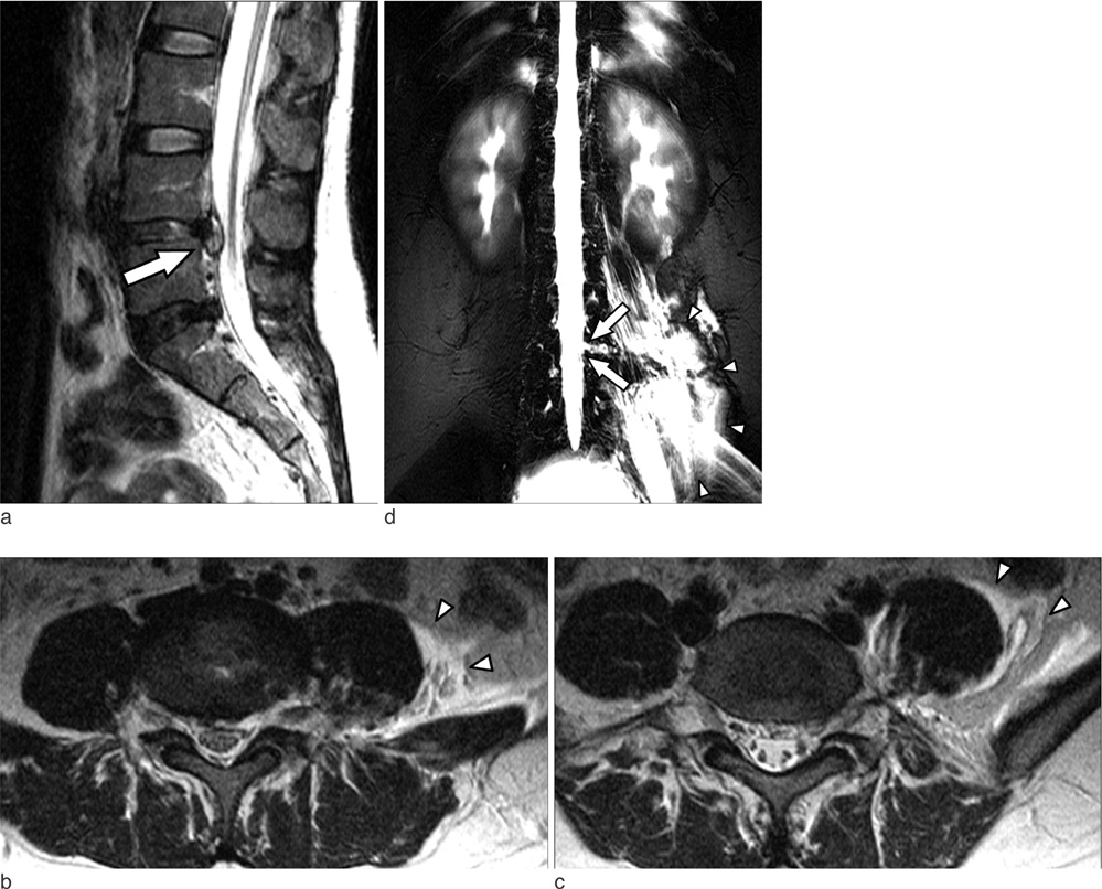
Fig. 1 Patient 1, a 28-year-old woman who developed orthostatic headache after endoscopic lumbar discectomy at the L4-5 level. a. T2-weighted sagittal image shows a focal hyperintense lesion (arrow) at the discectomy site at L4-5, with most changes being postoperative. b, c. L4-5, L5-S1 level axial T2-weighted images show left paravertebral fluid collection (arrowheads) but fail to visualize exact leakage site. d. 2D TSE coronal MR myelography shows the CSF leakage site (arrows) at the left side of the L4 level with paravertebral fluid collection (arrowheads) along the left paravertebral muscles.
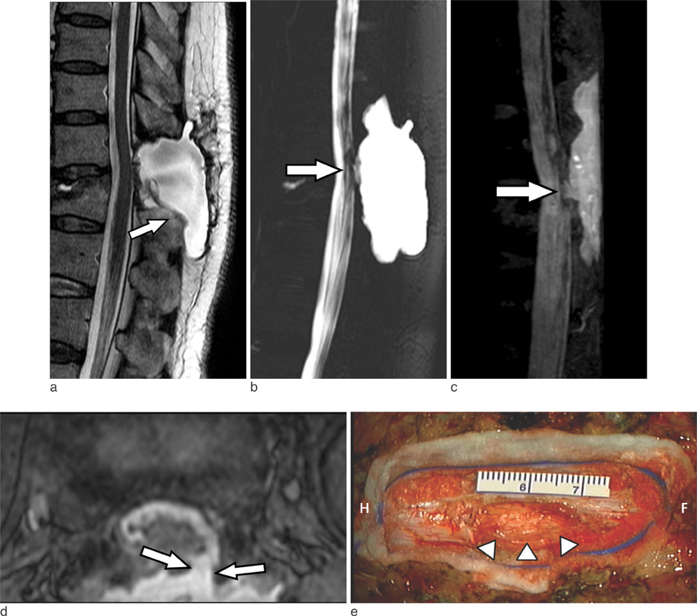
Fig. 2 Patient 2, a 64-year-old woman with orthostatic headache after resection of an intradural meningioma. (a) Postoperative T2-weighted sagittal image shows extradural fluid collection (arrow), but not the exact site of communication. 2D TSE sagittal (b) and MIP image of 3D BTFE MR myelography (c) show communication at the T12 level (arrow). (d) Axial reconstructed 3D BTFE image showing the defect on the left side of the thecal sac. (e) Intraoperative photograph of patient 2 shows that the dural defect (arrowheads) was located on the left side of dural sac and measured about 20 mm in length. The spinal cord is visible through the defect (Direction; H: head, F: foot).
Reference
-
1. Sin AH, Caldito G, Smith D, Rashidi M, Willis B, Nanda A. Predictive factors for dural tear and cerebrospinal fluid leakage in patients undergoing lumbar surgery. J Neurosurg Spine. 2006; 5:224–227.2. Jones AA, Stambough JL, Balderston RA, Rothman RH, Booth RE Jr. Long-term results of lumbar spine surgery complicated by unintended incidental durotomy. Spine. 1989; 14:443–446.3. Shapiro SA, Scully T. Closed continuous drainage of cerebrospinal fluid via a lumbar subarachnoid catheter for treatment or prevention of cranial/spinal cerebrospinal fluid fistula. Neurosurgery. 1992; 30:241–245.4. Eismont FJ, Wiesel SW, Rothman RH. Treatment of dural tears associated with spinal surgery. J Bone Joint Surg Am. 1981; 63:1132–1136.5. Lauer KK, Haddox JD. Epidural blood patch as treatment for a surgical durocutaneous fistula. J Clin Anesth. 1992; 4:45–47.6. El Gammal T, Sobol W, Wadlington VR, et al. Cerebrospinal fluid fistula: detection with MR cisternography. AJNR Am J Neuroradiol. 1998; 19:627–631.7. Rabin BM, Roychowdhury S, Meyer JR, Cohen BA, LaPat KD, Russell EJ. Spontaneous intracranial hypotension: spinal MR findings. AJNR Am J Neuroradiol. 1998; 19:1034–1039.8. Yoo HM, Kim SJ, Choi CG, et al. Detection of CSF leak in spinal CSF leak syndrome using MR myelography: correlation with radioisotope cisternography. AJNR Am J Neuroradiol. 2008; 29:649–654.9. Katramados A, Patel SC, Mitsias PD. Non-invasive magnetic resonance myelography in spontaneous intracranial hypotension. Cephalalgia. 2006; 26:1160–1164.10. Maycock NF, van Essen J, Pfitzner J. Post-laminectomy cerebrospinal fluid fistula treated with epidural blood patch. Spine. 1994; 19:2223–2225.11. Goldberg AL, Kershah SM. Advances in imaging of vertebral and spinal cord injury. J Spinal Cord Med. 2010; 33:105–116.12. Hashizume K, Watanabe K, Kawaguchi M, Fujiwara A, Furuya H. Comparison between computed tomography-myelography and radioisotope-cisternography findings in whiplash-associated disorders suspected to be caused by traumatic cerebrospinal fluid leak. Spine. 2012; 37:E721–E726.
- Full Text Links
-
- Actions
-
Cited
- CITED
-
- Close
- Share
-
- Similar articles
-
- Orthostatic Headache Diagnosed by Computed Tomography Myelography
- Subarachnoid Hemorrhage Presenting with Seizure due to Cerebrospinal Fluid Leakage after Spinal Surgery
- Spinal Presentation of Spontaneous Intracranial Hypotension
- Heavily T2-Weighted Magnetic Resonance Myelography as a Safe Cerebrospinal Fluid Leakage Detection Modality for Nontraumatic Subdural Hematoma
- A Case of Spontaneous Intracranial Hypotension: Detection of Cerebrospinal Fluid Leakage by Early Dynamic Radionuclide Cisternography
