J Korean Soc Magn Reson Med.
2012 Aug;16(2):184-188. 10.13104/jksmrm.2012.16.2.184.
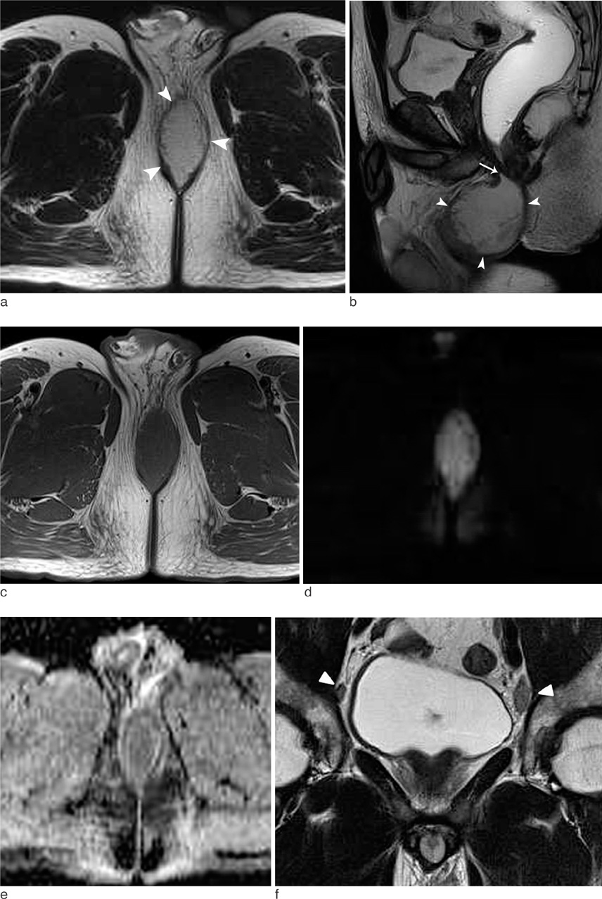
MR Imaging of Anoperineal Tuberculous Abscess: A Case Report
- Affiliations
-
- 1Department of Radiology, Anam Hospital, Korea University, College of Medicine, Korea. urorad@korea.ac.kr
- KMID: 2099854
- DOI: http://doi.org/10.13104/jksmrm.2012.16.2.184
Abstract
- Anoperineal tuberculosis is a rare extrapulmonary form of the disease and may present as abscess. We report a case of anoperineal tuberculous abscess, which showed low signal intensity on T1-weighted images, high signal intensity on T2-weighted images and diffusion restriction on diffusion weighted images.
MeSH Terms
Figure
Reference
-
1. Raviglione MC, Snider DE Jr, Kochi A. Global epidemiology of tuberculosis. Morbidity and mortality of a worldwide epidemic. JAMA. 1995. 273:220–226.2. Harland RW, Varkey B. Anal tuberculosis: report of two cases and literature review. Am J Gastroenterol. 1992. 87:1488–1491.3. Burrill J, Williams CJ, Bain G, Conder G, Hine AL, Misra RR. Tuberculosis: a radiologic review. Radiographics. 2007. 27:1255–1273.4. Candela F, Serrano P, Arriero JM, Teruel A, Reyes D, Calpena R. Perianal disease of tuberculous origin: report of a case and review of the literature. Dis Colon Rectum. 1999. 42:110–112.5. Yaghoobi R, Khazanee A, Bagherani N, Tajalli M. Gastrointestinal tuberculosis with anal and perianal involvement misdiagnosed as Crohn's disease for 15 years. Acta Derm Venereol. 2011. 91:348–349.6. Jinkins JR, Gupta R, Chang KH, Rodriguez-Carbajal J. MR imaging of central nervous system tuberculosis. Radiol Clin North Am. 1995. 33:771–786.7. Murata Y, Yamada I, Sumiya Y, Shichijo Y, Suzuki Y. Abdominal macronodular tuberculomas: MR findings. J Comput Assist Tomogr. 1996. 20:643–646.8. Morita S, Higuchi M, Takahata T, et al. Magnetic resonance imaging for multiple macronodular localized splenic tuberculosis. Clin Imaging. 2007. 31:134–136.9. Luthra G, Parihar A, Nath K, et al. Comparative evaluation of fungal, tubercular, and pyogenic brain abscesses with conventional and diffusion MR imaging and proton MR spectroscopy. AJNR Am J Neuroradiol. 2007. 28:1332–1338.10. Tappouni RF, Sarwani NI, Tice JG, Chamarthi S. Imaging of unusual perineal masses. AJR Am J Roentgenol. 2011. 196:W412–W420.


