J Korean Soc Radiol.
2015 Jun;72(6):431-434. 10.3348/jksr.2015.72.6.431.
Epidermoid Cyst of the Ovary in Young Woman: A Case Report
- Affiliations
-
- 1Department of Radiology, Keimyung University School of Medicine, Dongsan Medical Center, Daegu, Korea. kseehdr@dsmc.or.kr
- 2Department of Pathology, Keimyung University School of Medicine, Dongsan Medical Center, Daegu, Korea.
- KMID: 2098025
- DOI: http://doi.org/10.3348/jksr.2015.72.6.431
Abstract
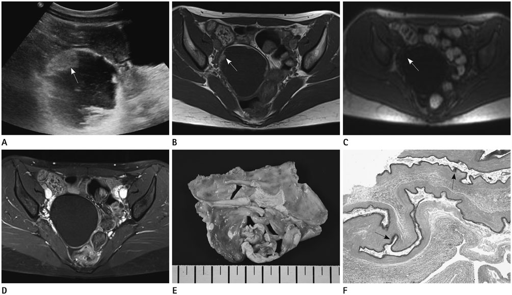
- In general, ovarian epidermoid cysts coexist with surface epithelial ovarian tumors. Pure epidermoid cysts are extremely rare diseases, comprising less than 1% of surface ovarian tumors. We present here a pathologically proven epidermoid cyst of the ovary in a young woman with ultrasonographic and magnetic resonance findings.
Figure
Reference
-
1. Khedmati F, Chirolas C, Seidman JD. Ovarian and paraovarian squamous-lined cysts (epidermoid cysts): a clinicopathologic study of 18 cases with comparison to mature cystic teratomas. Int J Gynecol Pathol. 2009; 28:193–196.2. Idress R, Ahmad Z, Minhas K, Kayani N. Epidermoid cyst of the ovary. J Pak Med Assoc. 2007; 57:263–264.3. Hiremath R, Chandrashekarayya SH, Manswini Pol TJ, Anegundi KR. A rare case of a submental epidermoid cyst: a case report. J Clin Diagn Res. 2011; 5:1452–1453.4. Young RH, Prat J, Scully RE. Epidermoid cyst of the ovary. A report of three cases with comments on histogenesis. Am J Clin Pathol. 1980; 73:272–276.5. Fan LD, Zang HY, Zhang XS. Ovarian epidermoid cyst: report of eight cases. Int J Gynecol Pathol. 1996; 15:69–71.6. Shinya T, Joja I, Hashimura S, Hayashi H, Gobara H, Kato K, et al. Magnetic resonance imaging features of epidermoid cyst of the ovaries: magnetic resonance and computed tomography findings. J Comput Assist Tomogr. 2006; 30:906–909.7. Kondi-Pafiti A, Filippidou-Giannopoulou A, Papakonstantinou E, Iavazzo C, Grigoriadis C. Epidermoid or dermoid cysts of the ovary? Clinicopathological characteristics of 28 cases and a new pathologic classification of an old entity. Eur J Gynaecol Oncol. 2012; 33:617–619.


