Diagnostic Accuracy and Inter-Observer Agreement of Shoulder Magnetic Resonance Arthrography in the Detection of Labral Lesion and Assessment of Lesion Location
- Affiliations
-
- 1Department of Radiology, School of Medicine, Ewha Womans University, Mokdong Hospital, Seoul, Korea. mshjy@ewha.ac.kr
- 2Department of Radiology, Gangseo MizMedi Hospital, Seoul, Korea.
- 3Department of Orthopaedic Surgery, School of Medicine, Ewha Womans University, Mokdong Hospital, Seoul, Korea.
- KMID: 2097983
- DOI: http://doi.org/10.3348/jksr.2012.67.6.465
Abstract
- PURPOSE
To evaluate the diagnostic accuracy and inter-observer agreement of magnetic resonance (MR) arthrography in the detection of labral lesions by location and to describe useful MR imaging findings of labral tears.
MATERIALS AND METHODS
Sixty-eight patients who underwent both pre-operative MR arthrography and arthroscopy were included. The location of the labrum was classified into anterior (2-6 o'clock), superior (12-2 o'clock), and posterior (6-12 o'clock). Sensitivity, specificity, accuracy, and inter-observer agreement of MR arthrography for the diagnosis of labral lesions by location were calculated. Frequency of MR imaging findings such as detachment, high signal intensity cleft, contour change, absence, and signal change of the labrum by location were analyzed.
RESULTS
35 anterior, 44 superior and 15 posterior labral lesions were detected by arthroscopy. The corresponding sensitivities were 91.4%, 79.5%, and 40.0%, specificities were 90.9%, 20.8%, and 86.8%, accuracies were 91.2%, 58.8%, and 76.5%, and kappa values were 0.823, 0.252, and 0.394, for anterior, superior, posterior lesions, respectively. The most common MR imaging findings were detachment in 60.0% of anterior labrums, high signal intensity cleft in 52.3% of superior labrums, and normal in 60.0% of posterior labrums.
CONCLUSION
Diagnostic accuracy and inter-observer agreement of MR arthrography in the diagnosis of labral lesions are high in anterior labrums and low in superior or posterior labrums. The useful MR imaging findings of labral tears were different according to labral location.
MeSH Terms
Figure
-

Fig. 1 Transverse (A), oblique coronal (B), and oblique sagittal (C) fat-saturated T1-weighted magnetic resonance (MR) arthrogram of the left shoulder in a 20-year-old man demonstrate detachment and anterior displacement of the anterior labrum (black arrow) and high signal intensity cleft (arrowhead) in superior labrum. The MR result of the posterior labrum (white arrow) is discordant between readers which is interpreted as small with blunted margin by reader 1 and as normal by reader 2. On arthroscopy, superior and anterior labral tear is diagnosed from 10 o'clock to 6 o'clock and posterior labrum appears to be normal (not shown).
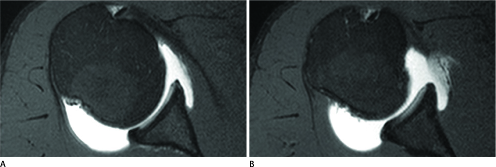
Fig. 2 A 19-year-old man with anterior labral tear (2-4 o'clock). Fat-saturated transverse T1-weighted images (A obtained cephalad to B) show absent anterior labrum with smooth glenoid margin.

Fig. 3 Magnetic resonance arthrograms from three different patients with or without superior labral lesions. A. A 36-year-old man with type II SLAP lesion confirmed with arthroscopy. Fat-saturated oblique coronal T1-weighted image shows laterally pointed, high signal intensity cleft from 10 to 12 o'clock (arrow), which was interpreted as tear by both readers. B. A 26-year-old man with type II SLAP lesion confirmed with arthroscopy. Fat-saturated oblique coronal T1-weighted image shows medially pointed, irregular high signal intensity at insertion site of biceps tendon (arrowhead), which was interpreted as normal by reader 1 and as tear by reader 2. C. A 19-year-old man without superior labral lesion by arthroscopy. Fat-saturated oblique coronal T1-weighted image show similar findings to A (arrowhead), which was interpreted as normal by reader 1 and as tear by reader 2. Note.-SLAP = superior labrum anterior and posterior tear
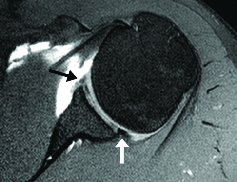
Fig. 4 A 28-year-old man with posterior and anterior labral tear (3-9 o'clock) confirmed with arthroscopy. Fat-saturated transverse T1-weighted image shows normal appeared posterior labrum (white arrow) by both readers. Concomitant anterior labral lesion is seen as increased signal intensity without poorly differentiated labrum (black arrow) because of synovial fibrous tissue and labral degeneration.
Reference
-
1. Dietrich TJ, Zanetti M, Saupe N, Pfirrmann CW, Fucentese SF, Hodler J. Articular cartilage and labral lesions of the glenohumeral joint: diagnostic performance of 3D water-excitation true FISP MR arthrography. Skeletal Radiol. 2010. 39:473–480.2. Song HT, Huh YM, Kim S, Lee SA, Kim SJ, Shin KH, et al. Anterior-inferior labral lesions of recurrent shoulder dislocation evaluated by MR arthrography in an adduction internal rotation (ADIR) position. J Magn Reson Imaging. 2006. 23:29–35.3. Song HT, Huh YM, Kim S, Kim SJ, Suh JS. The usefulness of virtual MR arthroscopy as an adjunct to conventional MR arthrography in detecting anterior labral lesions of the shoulder. AJR Am J Roentgenol. 2009. 192:W149–W155.4. Waldt S, Burkart A, Imhoff AB, Bruegel M, Rummeny EJ, Woertler K. Anterior shoulder instability: accuracy of MR arthrography in the classification of anteroinferior labroligamentous injuries. Radiology. 2005. 237:578–583.5. Chandnani VP, Yeager TD, DeBerardino T, Christensen K, Gagliardi JA, Heitz DR, et al. Glenoid labral tears: prospective evaluation with MRI imaging, MR arthrography, and CT arthrography. AJR Am J Roentgenol. 1993. 161:1229–1235.6. Jee WH, McCauley TR, Katz LD, Matheny JM, Ruwe PA, Daigneault JP. Superior labral anterior posterior (SLAP) lesions of the glenoid labrum: reliability and accuracy of MR arthrography for diagnosis. Radiology. 2001. 218:127–132.7. Amin MF, Youssef AO. The diagnostic value of magnetic resonance arthrography of the shoulder in detection and grading of SLAP lesions: comparison with arthroscopic findings. Eur J Radiol. 2012. 81:2343–2347.8. Palmer WE, Caslowitz PL. Anterior shoulder instability: diagnostic criteria determined from prospective analysis of 121 MR arthrograms. Radiology. 1995. 197:819–825.9. van Grinsven S, Kesselring FO, van Wassenaer-van Hall HN, Lindeboom R, Lucas C, van Loon CJ. MR arthrography of traumatic anterior shoulder lesions showed modest reproducibility and accuracy when evaluated under clinical circumstances. Arch Orthop Trauma Surg. 2007. 127:11–17.10. Holzapfel K, Waldt S, Bruegel M, Paul J, Heinrich P, Imhoff AB, et al. Inter- and intraobserver variability of MR arthrography in the detection and classification of superior labral anterior posterior (SLAP) lesions: evaluation in 78 cases with arthroscopic correlation. Eur Radiol. 2010. 20:666–673.11. Theodoropoulos JS, Andreisek G, Harvey EJ, Wolin P. Magnetic resonance imaging and magnetic resonance arthrography of the shoulder: dependence on the level of training of the performing radiologist for diagnostic accuracy. Skeletal Radiol. 2010. 39:661–667.12. Halma JJ, Eshuis R, Krebbers YM, Weits T, de Gast A. Interdisciplinary inter-observer agreement and accuracy of MR imaging of the shoulder with arthroscopic correlation. Arch Orthop Trauma Surg. 2012. 132:311–320.13. Bankart AS. Recurrent or habitual dislocation of the shoulder-joint. Br Med J. 1923. 15:1132–1133.14. Neviaser TJ. The anterior labroligamentous periosteal sleeve avulsion lesion: a cause of anterior instability of the shoulder. Arthroscopy. 1993. 9:17–21.15. Perthes G. Ueber operationen bei habitueller Schulterluxationen. Dtsch Z Chir. 1906. 85:199–227.16. Ryu JK, Yoon YC, Ryu KN, Rhee YG. Anterior labral tear: diagnostic value of MR arthrography of the shoulder. J Korean Radiol Soc. 2001. 45:61–67.17. Jin W, Ryu KN, Kwon SH, Rhee YG, Yang DM. MR arthrography in the differential diagnosis of type II superior labral anteroposterior lesion and sublabral recess. AJR Am J Roentgenol. 2006. 187:887–893.18. Saupe N, White LM, Bleakney R, Schweitzer ME, Recht MP, Jost B, et al. Acute traumatic posterior shoulder dislocation: MR findings. Radiology. 2008. 248:185–193.19. Chiavaras MM, Harish S, Burr J. MR arthrographic assessment of suspected posteroinferior labral lesions using flexion, adduction, and internal rotation positioning of the arm: preliminary experience. Skeletal Radiol. 2010. 39:481–488.20. Saleem AM, Lee JK, Novak LM. Usefulness of the abduction and external rotation views in shoulder MR arthrography. AJR Am J Roentgenol. 2008. 191:1024–1030.21. Park YH, Lee JY, Moon SH, Mo JH, Yang BK, Hahn SH, et al. MR arthrography of the labral capsular ligamentous complex in the shoulder: imaging variations and pitfalls. AJR Am J Roentgenol. 2000. 175:667–672.22. Kwak JY, Ha DH, Kim JS, Lee YS. Comparison of shoulder positions at MR arthrography: change of labroligamentous complex shape and diagnosis of labral tears. J Korean Radiol Soc. 2001. 45:499–505.
- Full Text Links
-
- Actions
-
Cited
- CITED
-
- Close
- Share
-
- Similar articles
-
- Anterior Labral Tear: Diagnostic Value of MR Arthrography of the Shoulder
- Accuracy of Magnetic Resonance Imaging and Computed Tomography Arthrography in Diagnosing Acetabular Labral Tears and Chondral Lesions
- Comparison of Diagnostic Accuracy of 3.0-T MR Arthrography and CT Arthrography in Intraarticular Hip Pathology
- 16-Slice MDCT Arthrography of the Shoulder: Accuracy for Detection of Glenoid Labral and Rotator Cuff Tears
- C. T. arthrography on Bankart lesion
