Giant Chest Wall Hematoma Mimicking Elastofibroma Dorsi: A Case Report
- Affiliations
-
- 1Department of Radiology, Daejeon St. Mary's Hospital, The Catholic University of Korea, Daejeon, Korea.
- 2Department of Thoracic Surgery, Daejeon St. Mary's Hospital, The Catholic University of Korea, Daejeon, Korea.
- 3Department of Pathology, Daejeon St. Mary's Hospital, The Catholic University of Korea, Daejeon, Korea.
- 4Department of Rehabilitation Medicine, Daejeon St. Mary's Hospital, The Catholic University of Korea, Daejeon, Korea. ces612@nate.com
- KMID: 2097931
- DOI: http://doi.org/10.3348/jksr.2011.64.2.151
Abstract
- Hematoma on the thoracic wall is very rare. We describe here a 63-year-old man with a huge chest wall hematoma and the man had no history of trauma. The patient was found to have a large mass located subjacent to the inferior angle of the right scapula area and the CT and MRI findings were similar to those of an elastofibroma dorsi. We describe the CT and MRI findings of this hematoma and how to make the differential diagnosis from elastofibroma dorsi.
MeSH Terms
Figure
-

Fig. 1 A. The axial noncontrast chest CT scan shows a poorly defined soft tissue mass (arrows) on the posterolateral aspect of the chest wall with an attenuation value similar to that of the neighboring muscle and suspicious linear interspersed hypodense areas. B. The axial postcontrast CT demonstrates no remarkable enhancement of the mass (arrows).
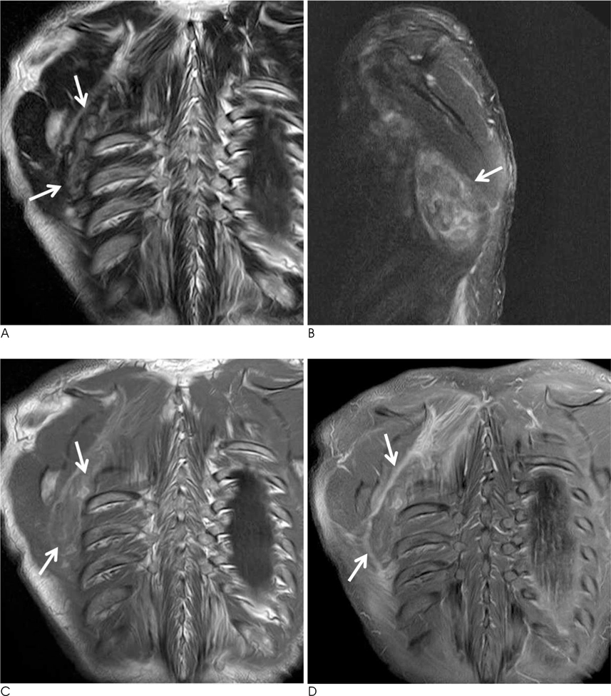
Fig. 2 A. The coronal T2-weighted MR image shows the soft tissue mass in the right subscapular region with heterogeneous signal intensity that included low signal intensity and high signal intensity linear strands. There is an associated peripheral dark signal rim (arrows). B. The fat suppressed sagittal T2-weighted MR image reveals heterogeneous hyperintensity (arrow). C. The coronal T1-weighted MR image shows an intermediate signal intensity mass lesion (arrow) in the right chest wall, with interspersed lines of high signal. D. The fat suppressed coronal T1-weighted MR image after contrast injection shows peripheral rim enhancement without central enhancement (arrow).

Fig. 3 The microscopic findings show a mixture of fibrin, several hemosiderin-laden macrophages and blood clots. Neither elastofibroma nor a fat component was present (H & E ×200).
Reference
-
1. O'Sullivan P, O'Dwyer H, Flint J, Munk PL, Muller N. Soft tissue tumours and mass-like lesions of the chest wall: a pictorial review of CT and MR findings. Br J Radiol. 2007; 80:574–580.2. Mike M, Kimura K, Watanabe J, Sasaki S, Kiyosawa Y, Momiyama H, et al. Giant hematoma on the thoracic wall: report of two cases. Surgery. 1999; 126:975–976.3. Kwon YS, Koh WJ, Kim TS, Lee KS, Kim BT, Shim YM. Chronic expanding hematoma of the thorax. Yonsei Med J. 2007; 48:337–340.4. Takahama M, Yamamoto R, Nakajima R, Izumi N, Tada H. Extrathoracic protrusion of a chronic expanding hematoma in the chest mimicking a soft tissue tumor. Gen Thorac Cardiovasc Surg. 2010; 58:202–204.5. McKenzie G, Raby N, Ritchie D. Pictorial review: non-neoplastic soft-tissue masses. Br J Radiol. 2009; 82:775–785.6. Lim HJ, Kim WT, Cho YK, Kim YJ. Radiologic findigs of bilateral elastofibroma dorsi: a case report. J Korean Radiol Soc. 2007; 56:283–287.7. Malghem J, Baudrez V, Lecouvet F, Lebon C, Maldague B, Vande Berg B. Imaging study findings in elastofibroma dorsi. Joint Bone Spine. 2004; 71:536–541.8. Reid JD, Kommareddi S. Chronic expanding hematomas: a clinicopathologic entity. JAMA. 1980; 244:2441–2442.9. Liu PT, Leslie KO, Beauchamp CP, Cherian SF. Chronic expanding hematoma of the thigh simulating neoplasm on gadolinium-enhanced MRI. Skeletal Radiol. 2006; 35:254–257.10. Burgener FA, Meyers SP, Tan RK, Zaunbauer W. Differntial diagnosis in magnetic resonance imaging. New York: Thieme;2002. p. 340–341.
- Full Text Links
-
- Actions
-
Cited
- CITED
-
- Close
- Share
-
- Similar articles
-
- Sonographic Findings of Bilateral Elastofibroma Dorsi: A Case Report
- Radiologic Findings of Bilateral Elastofibroma Dorsi: A Case Report
- Bilateral Elastofibroma Dorsi of the Shoulder: Case Report
- Bilateral Elastofibroma Dorsi in the Infrascapular Region: A case report
- Gant Infrascapular Rheumatoid Nodules Mimicking Elastofibroma Dorsi: A Case Report
