J Korean Soc Spine Surg.
2014 Dec;21(4):179-182. 10.4184/jkss.2014.21.4.179.
Old and New Fashion: Minimally Invasive Spine Surgery for Adjacent Segmental Spinal Stenosis after Luque Sublaminar Wiring and Posterolateral Fusion: Case Report
- Affiliations
-
- 1Department of Orthopedic Surgery, Sung-Ae Hospital, Seoul, Republic of Korea.
- 2Department of Orthopedic Surgery, College of Medicine, Hanyang University, Seoul, Republic of Korea. cnkang65@hanyang.ac.kr
- KMID: 2097869
- DOI: http://doi.org/10.4184/jkss.2014.21.4.179
Abstract
- STUDY DESIGN: A case report.
OBJECTIVES
To report a rare case of minimally invasive spine surgery after sublaminar wiring fixation with Luque rods. SUMMARY OF LITERATURE REVIEW: In the past, sublaminar wiring fixation with Luque rods was believed to be an effective fixation method; however, the development of transpedicular fixation resulted in the discontinuation of this method. Currently, instead of classical surgery using a broad incision, minimally invasive spine surgery is performed, which has a multitude of advantages.
MATERIALS AND METHODS
Initially, the patient underwent Luque sublaminar wiring and posterolateral fusion. After 25 years, minimally invasive TLIF and percutaneous transpedicular fixation were performed for the adjacent segmental spinal stenosis.
RESULTS
A good bone union is developing with an excellent clinical outcome, 14 months after the surgery.
CONCLUSIONS
We report a very rare case of a patient who underwent Luque sublaminar wiring and then, 25 years later, had spine surgery with minimally invasive techniques.
Keyword
MeSH Terms
Figure
-

Fig. 1. Lumbar spine anteroposterior (A) and lateral (B) radiographs taken at 25 years after operation. There are endplate sclerosis and traction spurs at proximal L2-L3 level. T2 sagittal (C) and axial (D) MRI taken at 25 yeaers after operation show severe spinal stenosis of L2-L3 level and more aggravated than 10 years ago.
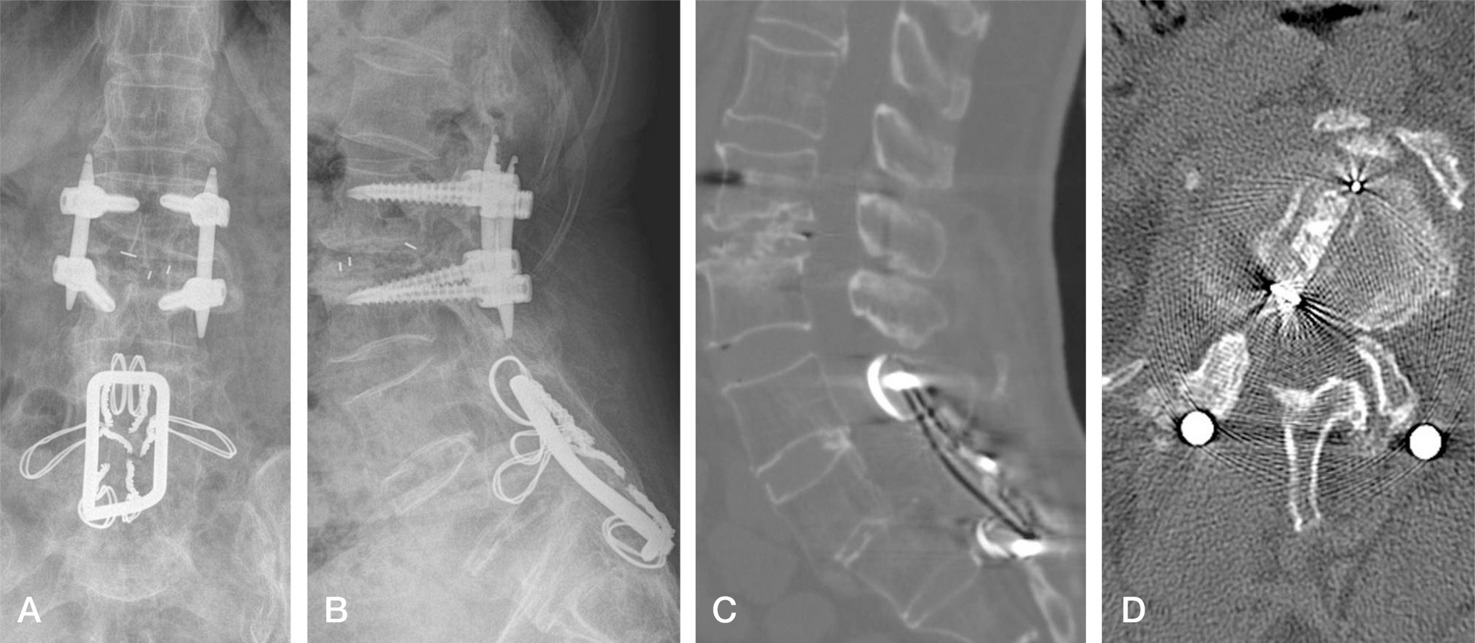
Fig. 2. Lumbar spine anteroposterior (A) and lateral (B) radiographs after minimally invasive TLIF and percutaneous transpedicular fixation were performed. Good bone union without loosening of screws is shown on sagittal (C) and axial (D) CT scan after 14 months of last surgery.
Reference
-
1. Pfirrmann CW, Metzdorf A, Zanetti M, Hodler J, Boos N. Magnetic resonance classification of lumbar intervertebral disc degeneration. Spine (Phila Pa 1976). 2001; 26:1873–8.
Article2. Foster MR. A functional classification of spinal instrumentation. Spine J. 2005; 5:682–94.
Article3. Luque ER, Cordosa A. Segmental correction of scoliosis with rigid internal fixation: preliminary report. Orthop Trans. 1977; 1:136–7.4. Luque ER. Segmental spinal instrumentation for the correction of scoliosis. Clin Orthop Relat Res. 1982; 163:192–8.5. Zdeblick TA, Becker PS, McAfee PC, Sutterlin CE, Coe JD, Gurr KR. Neuropathologic changes with experimental spinal instrumentation: transpedicular versus sublaminar fixation. J Spinal Disord. 1991; 4:221–8.6. Parsons JR, Chokshi BV, Lee CK, Gundlapalli RV, Stamer D. The biomechanical analysis of sublaminar wires and cables using luque segmental spinal instrumentation. Spine (Phila Pa 1976). 1997; 22:267–73.
Article7. Songer MN, Spencer DL, Meyer PR Jr, Jayaraman G. The use of sublaminar cables to replace Luque wires. Spine (Phila Pa 1976). 1991; Suppl:S418–21.
Article8. Zindrick MR, Wiltse LL, Widell EH, Thomas JC, Holland WR, Field BT, Spencer CW. A biomechanical study of in-trapeduncular screw fixation in the lumbosacral spine. Clin Orthop. 1986; 203:99–112.
Article9. Park Y, Ha JW. Comparison of one-level posterior lumbar interbody fusion performed with a minimally invasive approach or a traditional open approach. Spine (Phila Pa 1976). 2007; 32:537–43.
Article10. Tian NF, Wu YS, Zhang XL, Xu HZ, Chi YL, Mao FM. Minimally invasive versus open transforaminal lumbar interbody fusion: a meta-analysis based on the current evidence. Eur Spine J. 2013; 22:1741–9.
Article11. Chung HT, Na CO, Ha SH, Shin DR. Minimally invasive transforaminal lumbar interbody fusion. J Korean Soc Spine Surg. 2009; 16:24–9.
Article12. Park P, Garton HJ, Gala VC, Hoff JT, McGillicuddy JE. Adjacent segment disease after lumbar or lumbosacral fusion: review of the literature. Spine (Phila Pa 1976). 2004; 20:1938–44.
Article13. Yang JY, Lee JK, Song HS, Kim TH, Yeon KW. Correlation between clinical result and adjacent segment degeneration after lumbar spinal fusion. J Korean Soc Spine Surg. 2008; 15:38–43.
Article14. Cho JL, Park YS, Han JH, Lee CH, Roh WI. The change of adjacent segments after spinal fusion: Followup more than three years after spinal fusion. J Korean Soc Spine Surg. 1998; 5:239–46.
- Full Text Links
-
- Actions
-
Cited
- CITED
-
- Close
- Share
-
- Similar articles
-
- Technique of Safe Removal for Sublaminar Wiring
- Clinical Results of Segmental spinal instrumentation in Unstable Fracture and Fracture - Dislocation of the Thoracolumbar Spine
- Luque Instrumentation
- Segmental Spinal Instrumentation in the Treatment of Scoliosis
- Clinical Results of Segmental Spinal Instrumentation in Unstable Fracture and Fracture-Dislocation of the Thoracolumbar Spine
