Adventitial cystic disease of common femoral vein
- Affiliations
-
- 1Department of Surgery, Yeungnam University College of Medicine, Daegu, Korea. whkwun@med.yu.ac.kr
- KMID: 2096657
- DOI: http://doi.org/10.4174/jkss.2011.80.Suppl1.S75
Abstract
- Adventitial cystic disease (ACD) of venous system is an extremely rare condition. Very few reports of ACD in venous system have been described. In this report we discuss two cases of common femoral vein ACD that presented with a swollen leg by the obstruction of the vein. Ultrasound imaging showed the typical hypoechoic fluid filled cyst with a posterior acoustic window. Computed tomography scan and ascending venogram showed a stenosis to flow in the common femoral vein caused by an extrinsic mass. Trans-adventitial evacuation of cyst with removal of vein wall was performed for both cases. During operation we found the gelatinous material in the cysts arising in the wall of the common femoral vein and compressing the lumen. The patients were released after short hospitalization and have remained symptom free with no recurrence.
Keyword
MeSH Terms
Figure
-
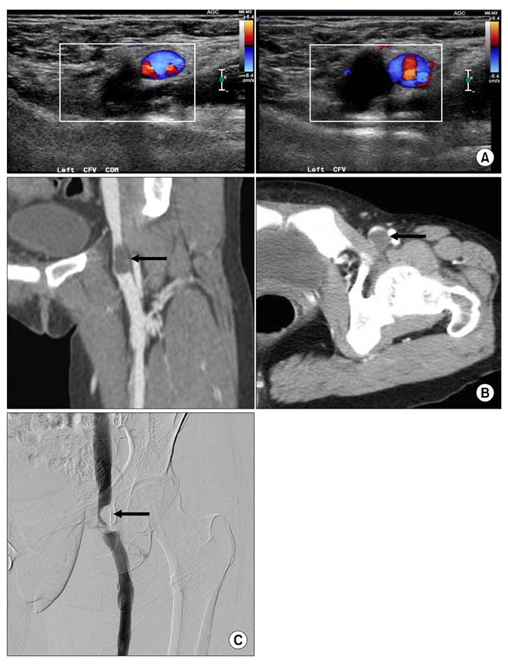
Fig. 1 Radiologic findings of 40-year-old woman. (A) Ultrasound imaging shows a typical hypoechoic fluid-filled cyst with a posterior acoustic window. Preoperative computed tomography (B) and ascending venogram (C) show focal filling defect in the left common femoral vein.
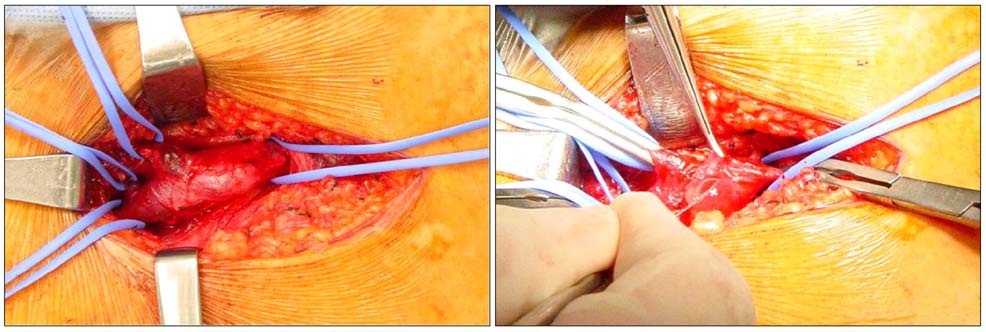
Fig. 2 Operative image shows the enlarged, obstructed common femoral vein with a bluish-colored mass visible through the wall of the vein and gelatinous material in large cyst arising in the posterior wall of the common femoral vein and compressing the lumen.
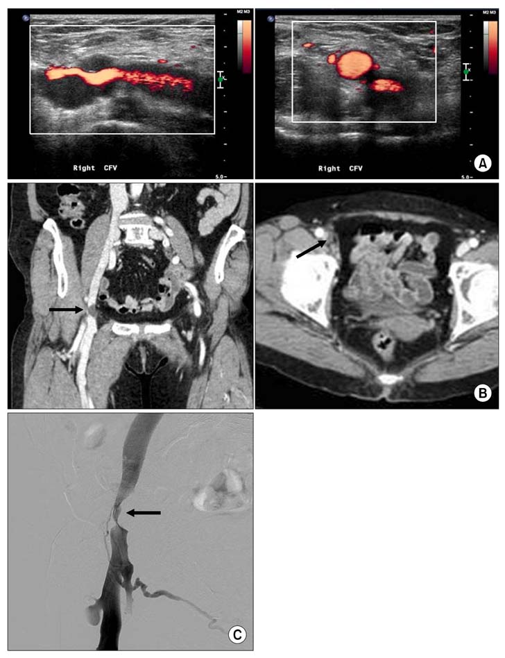
Fig. 3 Radiologic findings of 68-year-old woman. (A) Ultrasound imaging shows a typical hypoechoic fluid-filled cyst with a posterior acoustic window. Preoperative computed tomography (B) and ascending venogram (C) show focal obliteration of right common vein.

Fig. 4 Operative image shows the gelatinous material in large cyst arising in the lateral wall of the common femoral vein and compressing the lumen.
Reference
-
1. Sugimoto T, Yamamoto K, Tanaka S, Saitou N, Kikuchi C, Motohashi S, et al. Adventitial cystic disease of the femoral vein: report of a case. Surg Today. 2004. 34:286–288.2. Levien LJ, Benn CA. Adventitial cystic disease: a unifying hypothesis. J Vasc Surg. 1998. 28:193–205.3. Johnson JM, Kiankhooy A, Bertges DJ, Morris CS. Percutaneous image-guided aspiration and sclerosis of adventitial cystic disease of the femoral vein. Cardiovasc Intervent Radiol. 2009. 32:812–816.4. Mino MJ, Garrigues DG, Pierce DS, Arko FR. Cystic adventitial disease of the popliteal artery. J Vasc Surg. 2009. 49:1324.5. Dix FP, McDonald M, Obomighie J, Chalmers N, Thompson D, Benbow EW, et al. Cystic adventitial disease of the femoral vein presenting as deep vein thrombosis: a case report and review of the literature. J Vasc Surg. 2006. 44:871–874.6. Gasparis AP, Wall P, Ricotta JJ. Adventitial cystic disease of the external iliac vein presenting with deep venous thrombosis: a case report. Vasc Endovascular Surg. 2004. 38:273–276.
- Full Text Links
-
- Actions
-
Cited
- CITED
-
- Close
- Share
-
- Similar articles
-
- Adventitial Cystic Disease of the Common Femoral Vein Mimicking Deep Venous Thrombosis: A Case Report
- A Case of Adventitial Cystic Disease of the Femoral Vein
- Adventitial Cystic Disease of the Superficial Femoral Vein without a Joint Connection: A Case Report
- Adventitial Cystic Disease of the Common Femoral Artery: A Case Report and Literature Review
- Adventitial Cystic Disease of the Left External Iliac Vein: A Case Report
