Successful Percutaneous Coronary Intervention in an Anomalous Origin of the Right Coronary Artery From the Ascending Aorta Above the Left Sinus of the Valsalva
- Affiliations
-
- 1Department of Internal Medicine, Chungnam National University College of Medicine, Daejeon, Korea. jojeong@cnu.ac.kr
- 2Department of Radiology, Chungnam National University College of Medicine, Daejeon, Korea.
- KMID: 2094123
- DOI: http://doi.org/10.4070/kcj.2012.42.7.497
Abstract
- The anomalous origin of the right coronary artery (RCA) is a rare condition. Most RCA anomalies are usually found incidentally, but these findings have clinical significance because many patients, particularly young ones, present with sudden death, myocardial ischemia and syncope without other symptoms. We describe a case of a 39-year-old male patient that presented with effort chest pain and was diagnosed with anomalous RCA that originated from the ascending aorta with prior history of repairing ruptured sinus valsalva and ventricular septal defect. The anomalous origin of RCA was identified by multidetector computed tomography (MDCT). Successful percutaneous coronary intervention was performed guided by MDCT coronary images and intravascular ultrasound.
Keyword
MeSH Terms
Figure
-
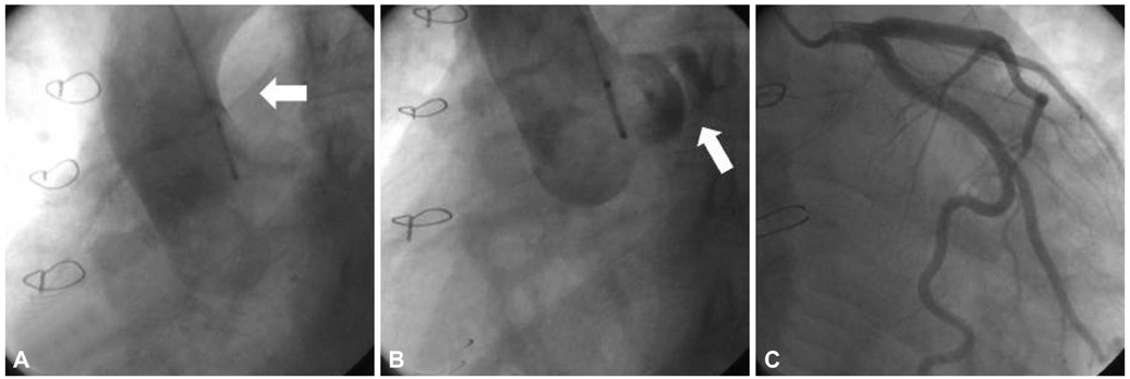
Fig. 1 Aortogram and coronary angiography. A: anomalous right coronary artery ostium (arrow). B: left main coronary artery originated from left coronary cusp (arrow). C: normal left coronary artery.

Fig. 2 MDCT coronary images. Volume rendering images and alternative view of coronary MDCT angiography showed ectopic ostium of RCA from the tubular portion of ascending aorta (A) and acute angulated tight stenosis (B). RCA originated from the left side and interposited between the pulmonary trunk and ascending aorta (C and D). MDCT: multidetector computed tomography, RCA: right coronary artery.

Fig. 3 Right coronary angiography. Baseline coronary angiogram showed an anomalous right coronary artery (RCA) ostium and subtotal occlusion of the proximal RCA (arrow).
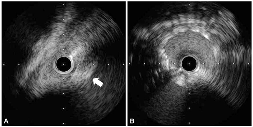
Fig. 4 The IVUS study. IVUS of the RCA. A: atherosclerotic plaque burdens on the proximal RCA (arrow). B: successfully implanted stent at the proximal RCA. IVUS: intravascular ultrasound, RCA: right coronary artery.
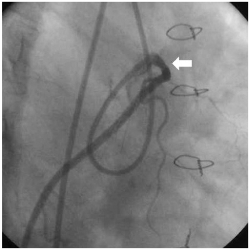
Fig. 5 Final right coronary angiography. The final coronary angiogram showed a successfully implanted stent at the proximal right coronary artery (arrow).
Reference
-
1. Kardos A, Babai L, Rudas L, et al. Epidemiology of congenital coronary artery anomalies: a coronary arteriography study on a central European population. Cathet Cardiovasc Diagn. 1997. 42:270–275.2. Yamanaka O, Hobbs RE. Coronary artery anomalies in 126,595 patients undergoing coronary arteriography. Cathet Cardiovasc Diagn. 1990. 21:28–40.3. Moreno R, Barrera-Ramirez C, Garcia J, Macaya C. Primary stenting of an anomalous left main trunk originating from the right coronary artery during acute myocardial infarction. J Invasive Cardiol. 2004. 16:159–161.4. Angelini P. Coronary artery anomalies--current clinical issues: definitions, classification, incidence, clinical relevance, and treatment guidelines. Tex Heart Inst J. 2002. 29:271–278.5. Hutchins GM, Miner MM, Boitnott JK. Vessel caliber and branch-angle of human coronary artery branch-points. Circ Res. 1976. 38:572–576.6. Zhang M, Kang WC, Ahn TH, Shin EK. Multidetector computed tomography for evaluation of ischemic etiology and a post-unroofing procedure for an anomalous origin of the right coronary artery from the left sinus of Valsalva. Korean Circ J. 2010. 40:251–252.7. Moon JY, Jeong HC, Cho JY, et al. Anomalous origin of a right coronary artery with extrinsic compression between the great vessels: the intravascular ultrasound images. Korean Circ J. 2008. 38:390–392.8. Sakakibara S, Konno S. Congenital aneurysm of the sinus of Valsalva: anatomy and classification. Am Heart J. 1962. 63:405–424.9. Scagliotti D, Fisher EA, Deal BJ, Gordon D, Chomka EV, Brundage BH. Congenital aneurysm of the left sinus of Valsalva with an aortopulmonary tunnel. J Am Coll Cardiol. 1986. 7:443–445.10. Angelini P, Velasco JA, Flamm S. Coronary anomalies: incidence, pathophysiology, and clinical relevance. Circulation. 2002. 105:2449–2454.11. Laurence L, Thurman R, Bruce TC. Atherosclerotic occlusions in anomalous left circumflex coronary arteries: a report of two unusual cases & a review of pertinent literature. Paroi Arterielle. 1975. 3:55–59.
- Full Text Links
-
- Actions
-
Cited
- CITED
-
- Close
- Share
-
- Similar articles
-
- Sudden Death Associated with Anomalous Left Coronary Artery Origin from Right Sinus of Valsalva with Posterior Course
- A Case of Acute Myocardial Infarction with the Anomalous Origin of the Right Coronary Artery from the Ascending Aorta above the Left Sinus of Valsalva and Left Coronary Artery from the Posterior Sinus of Valsalva
- Two Cases of Anomalous Origin of Coronary Artery
- Anomalous Origin of the Left Coronary Artery from the Right Sinus of Valsalva, which Presented as Acute Myocardial Infarction
- Unroofing Procedure in the Treatment of Anomalous Origin of Right Coronary Artery from Left Sinus of Valsalva between Aorta and Pulmonary Trunk
