Korean Circ J.
2012 Jul;42(7):487-491. 10.4070/kcj.2012.42.7.487.
Acute Coronary Stent Thrombosis in Cancer Patients: A Case Series Report
- Affiliations
-
- 1Division of Cardiology, Department of Internal Medicine, Seoul National University Hospital, Seoul, Korea.
- 2Division of Cardiology, Department of Internal Medicine, Seoul National University Bundang Hospital, Seongnam, Korea. changhwanyoon@gmail.com
- KMID: 2094121
- DOI: http://doi.org/10.4070/kcj.2012.42.7.487
Abstract
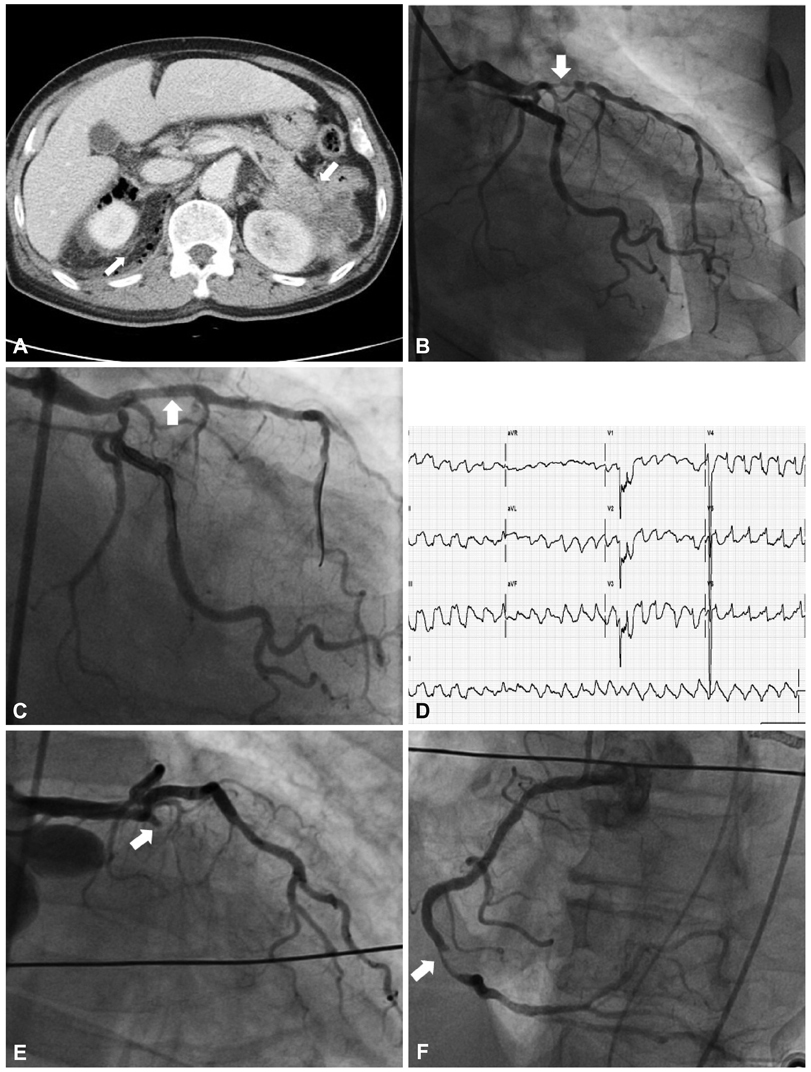
- There have been a growing numbers of patients diagnosed with malignancy and coronary artery disease simultaneously or serially. In the era of percutaneous coronary intervention (PCI), stent thrombosis has been a rare but challenging problem. Recently, we experienced two unique cases of acute stent thrombosis in patients with malignancy. The first case showed acute and subacute stent thrombosis after PCI. The second case revealed simultaneous thromboses in stent and non-treated native coronary artery. We believe that we need rigorous precautions in the treatment of patients with coronary artery disease and malignancy, especially with regards to deciding how and whether to revascularize, as well as which anti-platelet agents to select.
Keyword
MeSH Terms
Figure
Reference
-
1. Iakovou I, Schmidt T, Bonizzoni E, et al. Incidence, predictors, and outcome of thrombosis after successful implantation of drug-eluting st-ents. JAMA. 2005. 293:2126–2130.2. Blich M, Zeidan-Shwiri T, Petcherski S, Osherov A, Hammerman H. Incidence, predictors and outcome of drug-eluting stent thrombosis in real-world practice. J Invasive Cardiol. 2010. 22:461–464.3. Lee AY. Thrombosis in cancer: an update on prevention, treatment, and survival benefits of anticoagulants. Hematology Am Soc Hematol Educ Program. 2010. 2010:144–149.4. Khorana AA, Connolly GC. Assessing risk of venous thromboembolism in the patient with cancer. J Clin Oncol. 2009. 27:4839–4847.5. De Cicco M. The prothrombotic state in cancer: pathogenic mechanisms. Crit Rev Oncol Hematol. 2004. 50:187–196.6. Nakagawa T, Yasuno M, Tanahashi H, et al. A case of acute myocardial infarction: intracoronary thrombosis in two major coronary arteries due to hormone therapy. Angiology. 1994. 45:333–338.7. Canale ML, Camerini A, Stroppa S, et al. A case of acute myocardial infarction during 5-fluorouracil infusion. J Cardiovasc Med (Hagerstown). 2006. 7:835–837.8. Wijesinghe N, Thompson PI, McAlister H. Acute coronary syndrome induced by capecitabine therapy. Heart Lung Circ. 2006. 15:337–339.9. Kalapura T, Krishnamurthy M, Reddy CV. Acute myocardial infarction following gemcitabine therapy: a case report. Angiology. 1999. 50:1021–1025.10. Gemici G, Cinçin A, Değertekin M, Oktay A. Paclitaxel-induced ST-segment elevations. Clin Cardiol. 2009. 32:E94–E96.11. Lee SP, Suh JW, Park KW, et al. Study design and rationale of 'Influence of Cilostazol-based triple anti-platelet therapy on ischemic complication after drug-eluting stent implantation (CILON-T)' study: a multicenter randomized trial evaluating the efficacy of Cilostazol on ischemic vascular complications after drug-eluting stent implantation for coronary heart disease. Trials. 2010. 11:87.12. Gurbel PA, Bliden KP, Butler K, et al. Response to ticagrelor in clopidogrel nonresponders and responders and effect of switching therapies: the respond study. Circulation. 2010. 121:1188–1199.13. Montalescot G, Wiviott SD, Braunwald E, et al. Prasugrel compared with clopidogrel in patients undergoing percutaneous coronary intervention for ST-elevation myocardial infarction (TRITON-TIMI 38): double-blind, randomised controlled trial. Lancet. 2009. 373:723–731.
- Full Text Links
- Actions
-
Cited
- CITED
-
- Close
- Share
- Similar articles
-
- Very Late Stent Thrombosis in Coronary Bare-Metal Stent Implantation: A Case Report
- Simultaneous Multivessel Acute Stent Thrombosis in a Patient with Gastrointestinal Bleeding
- Recurrent Stent Thrombosis in Different Coronary Arteries
- Acute Stent Thrombosis after Coronary Stenting in Patients with Acute Coronary Syndrome
- Drug-Eluting Stent Used to Treat a Case of Recurrent Right Coronary Artery In-Stent Restenoses often Accompanied by Acute Inferior Wall Myocardial Infarction


