Segmental Tissue Doppler Image-Derived Tei Index in Patients With Regional Wall Motion Abnormalities
- Affiliations
-
- 1Department of Cardiology, Dong-A University College of Medicine, Busan, Korea. thpark65@dau.ac.kr
- KMID: 2094070
- DOI: http://doi.org/10.4070/kcj.2010.40.3.114
Abstract
- BACKGROUND AND OBJECTIVES
Although the Tei index is a useful predictor of global ventricular function, it has not been investigated at the level of regional myocardial function. We therefore investigated the segmental tissue Doppler image derived-Tei index (TDI-Tei index) in patients with regional wall motion abnormalities. SUBJECTS AND METHODS: We prospectively studied 17 patients (mean age 62+/-9 years, 5 women) with left ventricular (LV) regional wall motion abnormalities. The Tei index, defined as the sum of isovolumetric contraction time (IVCT) and isovolumetric relaxation time (IVRT) divided by ejection time (ET), was measured in the basal and mid segments of the LV walls from standard apical views (4-, 2-, and 5-chamber views). We also obtained TDI velocity data in each segment. LV wall motion was classified as normal, hypokinetic, or akinetic, based on visual analysis. The TDI-Tei index, peak systolic myocardial velocity (Sm), early diastolic myocardial velocity (Em), and late diastolic myocardial velocity (Am) were analyzed in a total of 203 segments. RESULTS: Mean LV ejection fraction was 41.8+/-8.5%. TDI-Tei indices of dysfunctional segments (akinesis or hypokinesis, n=63) were significantly higher than those of normal segments (n=140) (0.714+/-0.169 vs. 0.669+/-0.135, p=0.041, respectively). Average values of TDI-Tei index, Sm, Em, and Am were 0.742+/-0.201, 4.206+/-1.336, 5.258+/-1.867, and 5.578+/-2.354 in akinetic segments; 0.677+/-0.101, 4.908+/-1.615, 5.369+/-2.121, and 5.542+/-2.492 in hypokinetic segments; and 0.669+/-0.135, 5.409+/-1.519, 6.108+/-2.356, and 6.719+/-2.466 in normal segments, respectively. A significant negative correlation was apparent between the TDI-Tei index and Sm (r=-0.302, p<0.001). CONCLUSION: These data suggest that the value of the segmental TDI-Tei index differs significantly according to regional function grade.
Figure
-
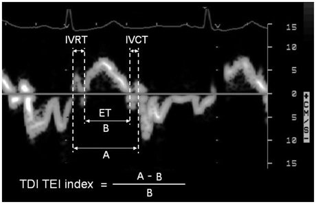
Fig. 1 A representative case of measurements for calculation of the TDI-Tei index. IVRT: isovolumetric relaxation time, IVCT: isovolumetric contraction time, ET: ejection time, TDI-Tei index: tissue Doppler image derived Tei index.
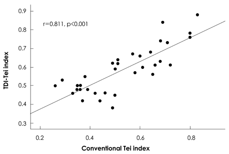
Fig. 2 Correlation of conventional Tei index and TDI-Tei index. TDI-Tei index: tissue Doppler image derived Tei index.
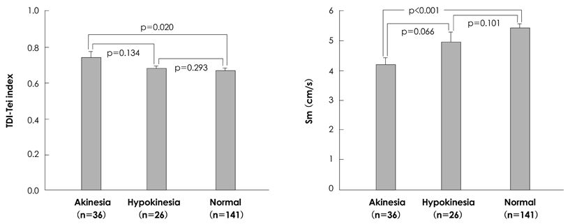
Fig. 3 Differences in TDI-Tei indices and peak systolic myocardial velocities among akinetic, hypokinetic, and normal myocardial segments. Sm: peak systolic myocardial velocity, TDI-Tei index: tissue Doppler image derived Tei index.
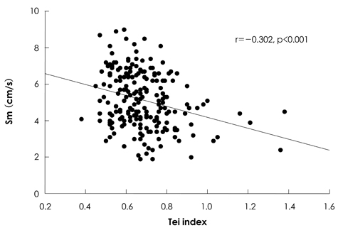
Fig. 4 Correlation of TDI-Tei index and peak systolic myocardial velocity. Sm: peak systolic myocardial velocity, TDI-Tei index: tissue Doppler image derived Tei index.
Reference
-
1. Tei C. New non-invasive index for combined systolic and diastolic ventricular function. J Cardiol. 1995. 26:135–136.2. Tei C, Ling LH, Hodge DO, et al. New index of combined systolic and diastolic myocardial performance: a simple and reproducible measure of cardiac function--a study in normals and dilated cardiomyopathy. J Cardiol. 1995. 26:357–366.3. Bruch C, Schmermund A, Marin D, et al. Tei-index in patients with mild-to-moderate congestive heart failure. Eur Heart J. 2000. 21:1888–1895.4. Møller JE, Egstrup K, Køber L, Poulsen SH, Nyvad O, Torp-Pedersen C. Prognostic importance of systolic and diastolic function after acute myocardial infarction. Am Heart J. 2003. 145:147–153.5. Abd El Rahman MY, Hui W, Dsebissowa F, et al. Comparison of the tissue Doppler-derived left ventricular Tei index to that obtained by pulse Doppler in patients with congenital and acquired heart disease. Pediatr Cardiol. 2005. 26:391–395.6. Su HM, Lin TH, Voon WC, et al. Differentiation of left ventricular diastolic dysfunction, identification of pseudonormal/restrictive mitral inflow pattern and determination of left ventricular filling pressure by Tei index obtained from tissue Doppler echocardiography. Echocardiography. 2006. 23:287–294.7. Sohn IS, Hang HS, Kim SJ, et al. Tissue Doppler image-derived myocardial performance (Tei Index) as a simple assessment of global cardiac function in adults. Korean Circ J. 2005. 35:315–321.8. Su HM, Lin TH, Voon WC, et al. Correlation of Tei index obtained from tissue Doppler echocardiography with invasive measurements of left ventricular performance. Echocardiography. 2007. 24:252–257.9. Lang RM, Bierig M, Devereux RB, et al. Recommendations for chamber quantification: a report from the American Society of Echocardiography's Guidelines and Standards Committee and the Chamber Quantification Writing Group, developed in conjunction with the European Association of Echocardiography, a branch of the European Society of Cardiology. J Am Soc Echocardiogr. 2005. 18:1440–1463.10. Tei C, Dujardin KS, Hodge DO, Kyle RA, Tajik AJ, Seward JB. Doppler index combining systolic and diastolic myocardial performance: clinical value in cardiac amyloidosis. J Am Coll Cardiol. 1996. 28:658–664.11. Dujardin KS, Tei C, Yeo TC, Hodge DO, Rossi A, Seward JB. Prognostic value of a Doppler index combining systolic and diastolic performance in idiopathic-dilated cardiomyopathy. Am J Cardiol. 1998. 82:1071–1076.12. Møller JE, Egstrup K, Køber L, Poulsen SH, Nyvad O, Torp-Pedersen C. Prognostic importance of systolic and diastolic function after acute myocardial infarction. Am Heart J. 2003. 145:147–153.13. Uzunhasan I, Bader K, Okcun B, Hatemi AC, Mutlu H. Correlation of the Tei index with left ventricular dilatation and mortality in patients with acute myocardial infarction. Int Heart J. 2006. 47:331–342.14. Sasao H, Noda R, Hasegawa T, Endo A, Oimatsu H, Takada T. Prognostic value of the Tei index combining systolic and diastolic myocardial performance in patients with acute myocardial infarction treated by successful primary angioplasty. Heart Vessels. 2004. 19:68–74.15. Baykan M, Erem C, Gedikli O, et al. Assessment of the Tei index by tissue Doppler imaging in patients with acromegaly: serum growth hormone level is associated with the Tei index. Echocardiography. 2008. 25:374–380.16. Harada K, Tamura M, Toyono M, Yasuoka K. Comparison of the right ventricular Tei index by tissue Doppler imaging to that obtained by pulsed Doppler in children without heart disease. Am J Cardiol. 2002. 90:566–569.
- Full Text Links
-
- Actions
-
Cited
- CITED
-
- Close
- Share
-
- Similar articles
-
- Tissue Doppler Image-Derived Myocardial Performance(Tei Index) as a Simple Assessment of Global Cardiac Function in Adults
- Modified Tei index in patients with Kawasaki disease by tissue doppler imaging
- The Evaluation of Myocardial Dyskinesia in the Patients with Coronary Artery Diseases
- Changes of Segmental Left Ventricular Wall Motion after PTCA in Ischemic Heart Diease
- Doppler Tei Index for Assessment of Subclinical Right Ventricular Dysfunction Associated with Inferior Wall Acute Myocardial Infarction
