Miniscrews versus surgical archwires for intermaxillary fixation in adults after orthognathic surgery
- Affiliations
-
- 1Department of Orthodontics, Pusan National University Dental Hospital, School of Dentistry, Pusan National University, Yangsan, Korea. softid@pusan.ac.kr
- 2Department of Oral and Maxillofacial Surgery, Pusan National University Dental Hospital, School of Dentistry, Pusan National University, Yangsan, Korea.
- KMID: 2074817
- DOI: http://doi.org/10.4041/kjod.2015.45.1.3
Abstract
OBJECTIVE
We compared the skeletal and dental changes that resulted from the use of two methods of intermaxillary fixation (IMF)-miniscrews and surgical archwire-in 74 adult patients who had Class III malocclusion and were treated with the same orthognathic surgical procedure at a hospital in Korea.
METHODS
All the patients underwent Le Fort I osteotomy and bilateral sagittal split ramus osteotomy with rigid fixation. They were divided into two groups according to the type of IMF used-group 1 underwent surgical archwire fixation and group 2 underwent orthodontic miniscrew fixation. In a series of cephalograms for each patient, we compared vertical and horizontal tooth-position measurements: (a) immediately after surgery (T0), (b) 3 months after surgery (T1), and (c) 6 months after surgery (T2). Cephalometric changes within each group were examined using one-way analysis of variance (ANOVA) while the independent samples t-test procedure was used to compare the two groups.
RESULTS
After surgery, the maxillary incisors tended to be proclined in both groups although there were no significant differences. Incisor overbite increased significantly in both groups from T0 to T1, and the miniscrew group (group 2) showed slightly greater overbite than the archwire group (group 1).
CONCLUSIONS
This study suggest that the use of orthodontic miniscrews and orthodontic surgical archwire for IMF in adult patients results in similar skeletal and dental changes.
Keyword
MeSH Terms
Figure
-
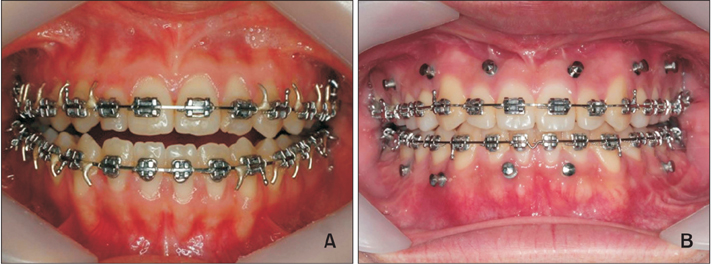
Figure 1 Intermaxillary fixation types. (A) Surgical archwire fixation and (B) Orthodontic miniscrew fixation.
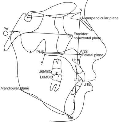
Figure 2 Landmarks and reference planes. S, Sella; N, Nasion; Po, Porion; Or, Orbitale; ANS, anterior nasal spine; PNS, posterior nasal spine; Me, Menton; U1E, incisal edge of maxillary central incisor (U1); U1A, root apex of U1; L1E, incisal edge of mandibular incisor (L1), L1A: root apex of L1; U6MBC, mesiobuccal cusp tip (MBC) of maxillary first molar; L6MBC, MBC of mandibular first molar; Frankfort horizontal plane (FH plane), line from Po to Or; N-perpendicular plane (N-perpend plane), perpendicular line to FH plane passing through Nasion.
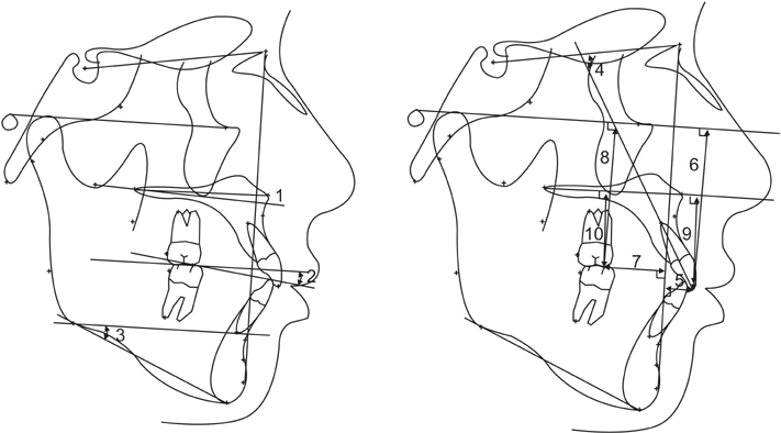
Figure 3 Cephalometric measurements. 1, FH-palatal plane; 2, FH-maxillary occlusal plane; 3, FMA; 4, U1 to SN; 5, U1-X; 6, U1-Y; 7, U6-X; 8, U6-Y; 9, U1-palatal; and 10, U6-palatal. Refer to Figure 2 and Table 1 for the definition of each measurements.
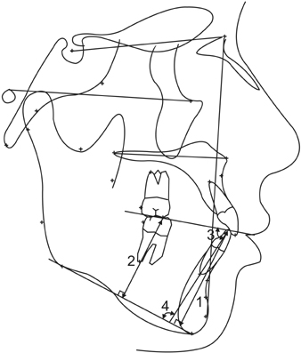
Figure 4 Cephalometric measurements of the mandible. 1, L1-mandibular plane; 2, L6-mandibular plane; 3, L1 to MnOP, and 4, IMPA. Refer to Figure 2 and Table 1 for the definition of each measurements.
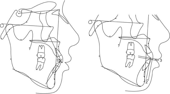
Figure 5 Cephalometric measurements of interdental relationship. 1, Interincisal angle; 2, incisor overjet; and 3, incisor overbite. Refer to Figure 2 and Table 1 for the definition of each measurements.
Cited by 1 articles
-
Comparison of postoperative changes in the distal and proximal segments between conventional and sliding mini-plate fixation following mandibular setback
Seong-Sik Kim, Kyoung-Ho Kwak, Ching-Chang Ko, Soo-Byung Park, Woo-Sung Son, Yong-Il Kim
Korean J Orthod. 2016;46(6):372-378. doi: 10.4041/kjod.2016.46.6.372.
Reference
-
1. Jones DC. The intermaxillary screw: a dedicated bicortical bone screw for temporary intermaxillary fixation. Br J Oral Maxillofac Surg. 1999; 37:115–116.
Article2. Wisth PJ, Isaksen TS. Changes in the vertical position of the anterior teeth after surgical correction of mandibular protrusion. Am J Orthod. 1980; 77:174–183.
Article3. Thota LG, Mitchell DA. Cortical bone screws for maxillomandibular fixation in orthognathic surgery. Br J Orthod. 1999; 26:325.
Article4. Arthur G, Berardo N. A simplified technique of maxillomandibular fixation. J Oral Maxillofac Surg. 1989; 47:1234.
Article5. Gibbons AJ, Hodder SC. A self-drilling intermaxillary fixation screw. Br J Oral Maxillofac Surg. 2003; 41:48–49.
Article6. Sahoo NK, Mohan R. IMF Screw: An ideal intermaxillary fixation device during open reduction of mandibular fracture. J Maxillofac Oral Surg. 2010; 9:170–172.
Article7. Lee JS, Park HS, Kyung HM. Micro-implant anchorage for lingual treatment of a skeletal Class II malocclusion. J Clin Orthod. 2001; 35:643–647.8. Park HS, Bae SM, Kyung HM, Sung JH. Micro-implant anchorage for treatment of skeletal Class I bialveolar protrusion. J Clin Orthod. 2001; 35:417–422.9. Park YC, Lee SY, Kim DH, Jee SH. Intrusion of posterior teeth using mini-screw implants. Am J Orthod Dentofacial Orthop. 2003; 123:690–694.
Article10. Ueki K, Marukawa K, Shimada M, Nakagawa K, Yamamoto E. The use of an intermaxillary fixation screw for mandibular setback surgery. J Oral Maxillofac Surg. 2007; 65:1562–1568.
Article11. Baek SH, Kim K, Choi JY. Evaluation of treatment modality for skeletal Class III malocclusion with labioversed upper incisors and/or protrusive maxilla: surgical movement and stability of rotational maxillary setback procedure. J Craniofac Surg. 2009; 20:2049–2054.
Article12. Hovell JH. Muscle patterning factors in the surgical correction of mandibular prognathism. J Oral Surg Anesth Hosp Dent Serv. 1964; 22:122–126.13. Troy BA, Shanker S, Fields HW, Vig K, Johnston W. Comparison of incisor inclination in patients with Class III malocclusion treated with orthognathic surgery or orthodontic camouflage. Am J Orthod Dentofacial Orthop. 2009; 135:146.e1–146.e9.
Article14. Lim LY, Cunningham SJ, Hunt NP. Stability of mandibular incisor decompensation in orthognathic patients. Int J Adult Orthodon Orthognath Surg. 1998; 13:189–199.15. Kim DK, Baek SH. Change in maxillary incisor inclination during surgical-orthodontic treatment of skeletal Class III malocclusion: comparison of extraction and nonextraction of the maxillary first premolars. Am J Orthod Dentofacial Orthop. 2013; 143:324–335.
Article16. Baek SH, Ahn HW, Kwon YH, Choi JY. Surgery-first approach in skeletal Class III malocclusion treated with 2-jaw surgery: evaluation of surgical movement and postoperative orthodontic treatment. J Craniofac Surg. 2010; 21:332–338.
Article17. Nagasaka H, Sugawara J, Kawamura H, Nanda R. "Surgery first" skeletal Class III correction using the Skeletal Anchorage System. J Clin Orthod. 2009; 43:97–105.18. Hashemi HM, Parhiz A. Complications using intermaxillary fixation screws. J Oral Maxillofac Surg. 2011; 69:1411–1414.
Article19. Coburn DG, Kennedy DW, Hodder SC. Complications with intermaxillary fixation screws in the management of fractured mandibles. Br J Oral Maxillofac Surg. 2002; 40:241–243.
Article20. Roccia F, Tavolaccini A, Dell'Acqua A, Fasolis M. An audit of mandibular fractures treated by intermaxillary fixation using intraoral cortical bone screws. J Craniomaxillofac Surg. 2005; 33:251–254.
Article21. Holmes S, Hutchison I. Caution in use of bicortical intermaxillary fixation screws. Br J Oral Maxillofac Surg. 2000; 38:574.
Article22. Choi JH, Yu HS, Lee KJ, Park YC. Three-dimensional evaluation of maxillary anterior alveolar bone for optimal placement of miniscrew implants. Korean J Orthod. 2014; 44:54–61.
Article23. Kim JH, Park YC. Evaluation of mandibular cortical bone thickness for placement of temporary anchorage devices (TADs). Korean J Orthod. 2012; 42:110–117.
Article24. Kim JS, Choi SH, Cha SK, Kim JH, Lee HJ, Yeom SS, et al. Comparison of success rates of orthodontic mini-screws by the insertion method. Korean J Orthod. 2012; 42:242–248.
Article
- Full Text Links
-
- Actions
-
Cited
- CITED
-
- Close
- Share
-
- Similar articles
-
- A study on positional changes of the teeth and mandible according to fixation type during intermaxillary fixation period after mandibular setback
- Clinical Experiences of New Intermaxillary Fixation Method without Tooth Ligation
- An Experimental Study Of Effect Of Intermaxillary Fixation And Occusal Splint On Pulmonary Function
- Two part mini-implant as an efficient tool for intermaxillary fixation
- A Clinical study on Pulmonary Function after Intermaxillary Fixation
