Korean J Urol.
2015 Jan;56(1):56-62. 10.4111/kju.2015.56.1.56.
Can stone density on plain radiography predict the outcome of extracorporeal shockwave lithotripsy for ureteral stones?
- Affiliations
-
- 1Department of Urology, Dongguk University Ilsan Hospital, Goyang, Korea. urmarine@dumc.or.kr
- 2Department of Radiology, Dongguk University Ilsan Hospital, Goyang, Korea.
- KMID: 2070269
- DOI: http://doi.org/10.4111/kju.2015.56.1.56
Abstract
- PURPOSE
The objective was to determine whether stone density on plain radiography (kidney-ureter-bladder, KUB) could predict the outcome of extracorporeal shockwave lithotripsy (ESWL) for ureteral stones.
MATERIALS AND METHODS
A total of 223 patients treated by ESWL for radio-opaque ureteral stones of 5 to 20 mm were included in this retrospective study. All patients underwent routine blood and urine analyses, plain radiography (KUB), and noncontrast computed tomography (NCCT) before ESWL. Demographic, stone, and radiological characteristics on KUB and NCCT were analyzed. The patients were categorized into two groups: lower-density (LD) group (radiodensity less than or equal to that of the 12th rib, n=163) and higher-density (HD) group (radiodensity greater than that of the 12th rib, n=60). Stone-free status was assessed by KUB every week after ESWL. A successful outcome was defined as stone free within 1 month after ESWL.
RESULTS
Mean stone size in the LD group was significantly smaller than that in the HD group (7.5+/-1.4 mm compared with 9.9+/-2.9 mm, p=0.002). The overall success rates in the LD and HD groups were 82.1% and 60.0%, respectively (p=0.007). The mean duration of stone-free status and average number of SWL sessions required for success in the two groups were 21.7 compared with 39.2 days and 1.8 compared with 2.3, respectively (p<0.05). On multivariate logistic analysis, stone size and time to ESWL since colic and radiodensity of the stone on KUB were independent predictors of successful ESWL.
CONCLUSIONS
Our data suggest that larger stone size, longer time to ESWL, and ureteral stones with a radiodensity greater than that of the 12th rib may be at a relatively higher risk of ESWL failure 1 month after the procedure.
Keyword
MeSH Terms
Figure
-
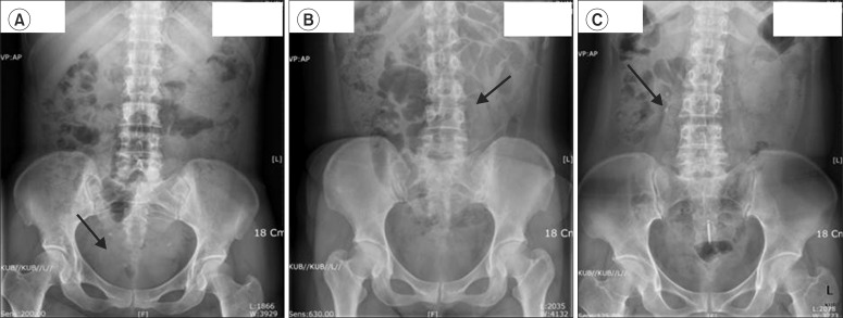
Fig. 1 Stone density classification compared with the density of the 12th rib: lower (arrow, A), equal (arrow, B), and higher density (arrow, C).
Cited by 1 articles
-
Impact of Pretreatment Hydronephrosis on the Success Rate of Shock Wave Lithotripsy in Patients with Ureteral Stone
Ki Don Chang, Joo Yong Lee, Sung Yoon Park, Dong Hyuk Kang, Hyung Ho Lee, Kang Su Cho
Yonsei Med J. 2017;58(5):1000-1005. doi: 10.3349/ymj.2017.58.5.1000.
Reference
-
1. Stamatelou KK, Francis ME, Jones CA, Nyberg LM, Curhan GC. Time trends in reported prevalence of kidney stones in the United States: 1976-1994. Kidney. 2003; 63:1817–1823.2. Tiselius HG, Ackermann D, Alken P, Buck C, Conort P, Gallucci M, et al. Guidelines on urolithiasis. Eur Urol. 2001; 40:362–371. PMID: 11713390.3. Tanaka M, Yokota E, Toyonaga Y, Shimizu F, Ishii Y, Fujime M, et al. Stone attenuation value and cross-sectional area on computed tomography predict the success of shock wave lithotripsy. Korean J Urol. 2013; 54:454–459. PMID: 23878688.
Article4. Pareek G, Hedican SP, Lee FT Jr, Nakada SY. Shock wave lithotripsy success determined by skin-to-stone distance on computed tomography. Urology. 2005; 66:941–944. PMID: 16286099.
Article5. Preminger GM, Tiselius HG, Assimos DG, Alken P, Buck AC, Gallucci M, et al. 2007 Guideline for the management of ureteral calculi. Eur Urol. 2007; 52:1610–1631. PMID: 18074433.
Article6. Dretler SP. Stone fragility: a new therapeutic distinction. J Urol. 1988; 139:1124–1127. PMID: 3361657.
Article7. Bon D, Dore B, Irani J, Marroncle M, Aubert J. Radiographic prognostic criteria for extracorporeal shock-wave lithotripsy: a study of 485 patients. Urology. 1996; 48:556–560. PMID: 8886060.
Article8. Choi HJ, Jung JH, Bae J, Cho MC, Lee HW, Lee KS. Usefulness of early extracorporeal shock wave lithotripsy in colic patients with ureteral stones. Korean J Urol. 2012; 53:853–859. PMID: 23301130.
Article9. Wang M, Shi Q, Wang X, Yang K, Yang R. Prediction of outcome of extracorporeal shock wave lithotripsy in the management of ureteric calculi. Urol Res. 2011; 39:51–57. PMID: 20401653.
Article10. Williams JC Jr, Saw KC, Paterson RF, Hatt EK, McAteer JA, Lingeman JE. Variability of renal stone fragility in shock wave lithotripsy. Urology. 2003; 61:1092–1096. PMID: 12809867.
Article11. Dretler SP, Polykoff G. Calcium oxalate stone morphology: fine tuning our therapeutic distinctions. J Urol. 1996; 155:828–833. PMID: 8583586.
Article12. Dretler SP, Spencer BA. CT and stone fragility. J Endourol. 2001; 15:31–36. PMID: 11248917.
Article13. Hidas G, Eliahou R, Duvdevani M, Coulon P, Lemaitre L, Gofrit ON, et al. Determination of renal stone composition with dual-energy CT: in vivo analysis and comparison with x-ray diffraction. Radiology. 2010; 257:394–401. PMID: 20807846.
Article14. Motley G, Dalrymple N, Keesling C, Fischer J, Harmon W. Hounsfield unit density in the determination of urinary stone composition. Urology. 2001; 58:170–173. PMID: 11489691.
Article15. Ng CF, Siu DY, Wong A, Goggins W, Chan ES, Wong KT. Development of a scoring system from noncontrast computerized tomography measurements to improve the selection of upper ureteral stone for extracorporeal shock wave lithotripsy. J Urol. 2009; 181:1151–1157. PMID: 19152949.
Article16. Shah K, Kurien A, Mishra S, Ganpule A, Muthu V, Sabnis RB, et al. Predicting effectiveness of extracorporeal shockwave lithotripsy by stone attenuation value. J Endourol. 2010; 24:1169–1173. PMID: 20575686.
Article17. Bandi G, Meiners RJ, Pickhardt PJ, Nakada SY. Stone measurement by volumetric three-dimensional computed tomography for predicting the outcome after extracorporeal shock wave lithotripsy. BJU Int. 2009; 103:524–528. PMID: 19007365.
Article18. Gupta NP, Ansari MS, Kesarvani P, Kapoor A, Mukhopadhyay S. Role of computed tomography with no contrast medium enhancement in predicting the outcome of extracorporeal shock wave lithotripsy for urinary calculi. BJU Int. 2005; 95:1285–1288. PMID: 15892818.
Article19. Yoshida S, Hayashi T, Ikeda J, Yoshinaga A, Ohno R, Ishii N, et al. Role of volume and attenuation value histogram of urinary stone on noncontrast helical computed tomography as predictor of fragility by extracorporeal shock wave lithotripsy. Urology. 2006; 68:33–37. PMID: 16806419.
Article
- Full Text Links
-
- Actions
-
Cited
- CITED
-
- Close
- Share
-
- Similar articles
-
- Non-surgical treatment of urinary stone
- The Comparison of Efficacy of Ureteroscopic Removal and Shockwave Lithotripsy in Lower Ureteral Stones
- Clinical experience of ureteral stones by extracorporeal shock wave lithotripsy
- Comparison between Rigid Ureteroscopic Stone Removal (URS) and Extracorporeal Shock Wave Lithotripsy (ESWL) for Large (>10mm) Upper Ureteral Stones
- The Effect of Tamsulosin and Nifedipine on Expulsion of Ureteral Stones after Extracorporeal Shock Wave Lithotripsy
