Real Stereopsis Test Using a Three-Dimensional Display with Tridef Software
- Affiliations
-
- 1Institute of Vision Research, Department of Ophthalmology, Yonsei University College of Medicine, Seoul, Korea. shhan222@yuhs.ac
- 2Department of Radiology, Yonsei University College of Medicine, Seoul, Korea.
- KMID: 2070218
- DOI: http://doi.org/10.3349/ymj.2014.55.6.1672
Abstract
- PURPOSE
To investigate horizontal image disparity in three-dimensional (3-D) perception using 3-D animations in normal control patients and patients with intermittent exotropia, anisometropic amblyopia, and partially accommodative esotropia.
MATERIALS AND METHODS
A total of 133 subjects were included. Stereopsis was measured using the Titmus Stereo test (Stereo Optical Inc., Chicago, IL, USA) and a 3-D stereopsis test with a 15 inch 3-D display laptop, adjusting 3-D parameters of 0 mm horizontal disparity to 15 mm horizontal disparity.
RESULTS
When compared with normal controls, the average threshold of the 3-D stereopsis test was significantly reduced for esotropia patients (p<0.001) and for anisometric amblyopia patients (p<0.001), compared to normal controls. No significant difference was observed between normal controls and intermittent exotropia patients (p=0.082). The 3-D stereopsis test was correlated with the Titmus Stereo test (Spearman's rho=0.690, p<0.001). Mean difference in stereoacuity was 1.323 log seconds of arc (95% limits of agreement: 0.486 to 2.112), and 125 (92.5%) patients were within the limits of agreement.
CONCLUSION
This study demonstrated that a 3-D stereopsis test with animation is highly correlated with the Titmus Stereo test; nevertheless, 3-D stereopsis with animations generates more image disparities than the conventional Titmus Stereo test. The 3-D stereopsis test is highly predictive for estimating real stereopsis in a 3-D movie theater.
MeSH Terms
Figure
-
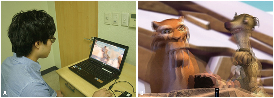
Fig. 1 (A) Subject watches the 3-dimensional (3-D) movie displayed by a 15 inch 3-D laptop computer. (B) The video clip was converted into a 3-D movie by a TriDef 3-D media player.
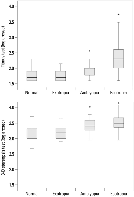
Fig. 2 Box plot illustrating the results of the Titmus Stereo and three-dimensional (3-D) stereopsis tests for each group of patients. *Statistical significance compared to normal controls. arcsec, seconds of arc.
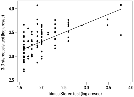
Fig. 3 Scatter plot showing the association between the Titmus Stereo and three-dimensional (3-D) stereopsis tests (Spearman's rho=0.690, p<0.001). arcsec, seconds of arc.
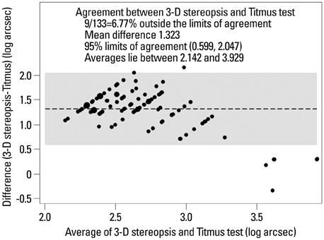
Fig. 4 Bland-Altman plot showing differences between the three-dimensional (3-D) stereopsis and Titmus Stereo tests for 133 subjects. Gray area shows 95% limits of agreement with 95% confidence intervals. Mean difference between the 3-D stereopsis and Titmus tests was 1.323 log arcsec. arcsec, seconds of arc.
Reference
-
1. Wardle SG, Gillam BJ. Phantom surfaces in da Vinci stereopsis. J Vis. 2013; 13:16.
Article2. Wallace DK, Lazar EL, Melia M, Birch EE, Holmes JM, Hopkins KB, et al. Stereoacuity in children with anisometropic amblyopia. J AAPOS. 2011; 15:455–461.
Article3. O'Connor AR, Fawcett SI, Stager DR, Birch EE. Factors influencing sensory outcome following surgical correction of infantile esotropia. Am Orthopt J. 2002; 52:69–74.4. Lee SY, Isenberg SJ. The relationship between stereopsis and visual acuity after occlusion therapy for amblyopia. Ophthalmology. 2003; 110:2088–2092.
Article5. Adams WE, Leske DA, Hatt SR, Mohney BG, Birch EE, Weakley DR Jr, et al. Improvement in distance stereoacuity following surgery for intermittent exotropia. J AAPOS. 2008; 12:141–144.
Article6. Birch EE, Wang J. Stereoacuity outcomes after treatment of infantile and accommodative esotropia. Optom Vis Sci. 2009; 86:647–652.
Article7. Cooper J. Clinical stereopsis testing: contour and random dot stereograms. J Am Optom Assoc. 1979; 50:41–46.8. Westheimer G. Clinical evaluation of stereopsis. Vision Res. 2013; 90:38–42.
Article9. Fawcett SL, Birch EE. Interobserver test-retest reliability of the Randot preschool stereoacuity test. J AAPOS. 2000; 4:354–358.
Article10. Adams WE, Leske DA, Hatt SR, Holmes JM. Defining real change in measures of stereoacuity. Ophthalmology. 2009; 116:281–285.
Article11. Birch EE. Marshall Parks lecture. Binocular sensory outcomes in accommodative ET. J AAPOS. 2003; 7:369–373.12. Stathacopoulos RA, Rosenbaum AL, Zanoni D, Stager DR, McCall LC, Ziffer AJ, et al. Distance stereoacuity. Assessing control in intermittent exotropia. Ophthalmology. 1993; 100:495–500.13. Fujikado T, Hosohata J, Ohmi G, Tano Y. A clinical evaluation of stereopsis required to see 3-D images. Ergonomics. 1996; 39:1315–1320.
Article14. Yang SN, Schlieski T, Selmins B, Cooper SC, Doherty RA, Corriveau PJ, et al. Stereoscopic viewing and reported perceived immersion and symptoms. Optom Vis Sci. 2012; 89:1068–1080.
Article15. Tomaç S, Altay Y. Near stereoacuity: development in preschool children; normative values and screening for binocular vision abnormalities; a study of 115 children. Binocul Vis Strabismus Q. 2000; 15:221–228.16. Köhler L, Stigmar G. Vision screening of four-year-old children. Acta Paediatr Scand. 1973; 62:17–27.
Article
- Full Text Links
-
- Actions
-
Cited
- CITED
-
- Close
- Share
-
- Similar articles
-
- Automated Techniques for the Sectioned Images of Visible Korean
- Change of Stereoacuity with Aging in Normal Eyes
- Evaluation of Criteria of Stereoacuity for Titmus, Randot & TNO Stereotests
- Regional Metabolic Changes Influencing Three-Dimensional Perception in Parkinson’s Disease
- The Post-operative Changes of Stereopsis in Adult Strabismus
