Renal Pelvic Urothelial Carcinoma With Vena Caval Thrombus Mimicking Renal Cell Carcinoma
- Affiliations
-
- 1Department of Urology, Urological Science Institute, Severance Hospital, Yonsei University College of Medicine, Seoul, Korea. youngd74@yuhs.ac
- 2Department of Pathology, Yonsei University College of Medicine, Seoul, Korea.
- 3Department of Urology, Urological Science Institute, Gangnam Severance Hospital, Yonsei University College of Medicine, Seoul, Korea.
- 4Clinical Trials Center for Medical Devices, Severance Hospital, Yonsei University College of Medicine, Seoul, Korea.
- KMID: 2069782
- DOI: http://doi.org/10.4111/kju.2014.55.9.624
Abstract
- A 61-year-old man presented with a right renal mass with a vena caval thrombus on computed tomography that was consistent with renal cell carcinoma. The results of routine laboratory examinations and urinalysis were within normal limits. Preoperative planning was critical owing to the presence of the vena caval thrombus. A radical nephrectomy, vena caval thrombectomy, and regional lymphadenectomy were done. The pathologic report was consistent with a high-grade, invasive urothelial carcinoma, with sarcomatoid differentiation involving the renal vein and inferior vena cava (Stage IV, T4N0M0). Thus, this was a rare case of upper tract urothelial carcinoma. Adjuvant chemotherapy with the methotrexate, vinblastine, doxorubicin, cisplatinum regimen is scheduled. To our knowledge, this is the first report in Korea of upper tract urothelial carcinoma of the sarcomatoid type with a vena caval thrombus.
MeSH Terms
Figure
-
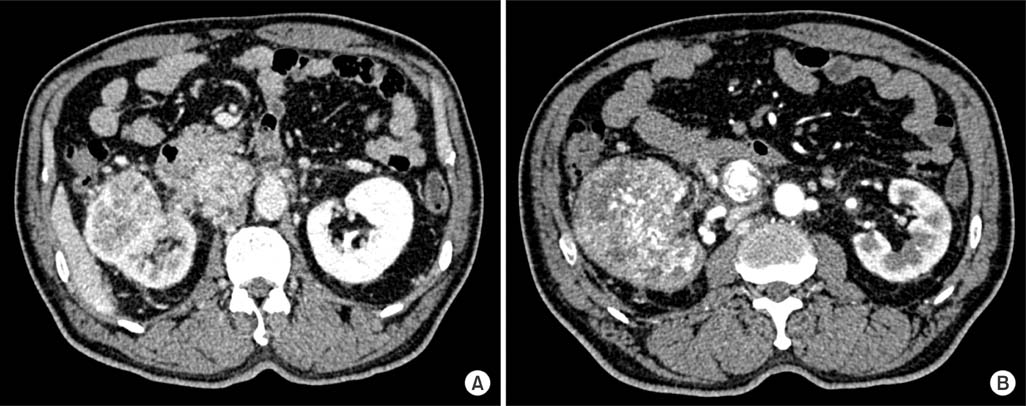
FIG. 1 Computed tomography revealed an approximately 10-cm-sized renal mass with partial necrosis (A) and invasion to the right renal vein and inferior vena cava (B).
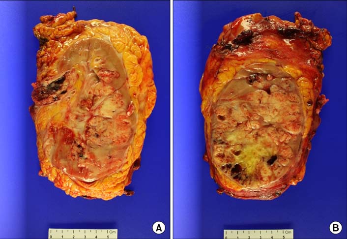
FIG. 2 Bisected kidney revealed an ill-defined, infiltrative, whitish soft mass (9.5 cm×7 cm), mainly involving the renal pelvis (A). The ureter was dilated owing to extension of the tumor (B).
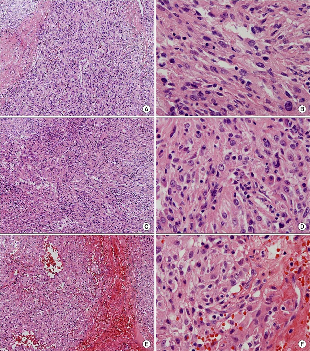
FIG. 3 Urothelial carcinoma, of a high grade, invaded the renal cortex limited to the capsule (A: H&E, ×100; B: H&E, ×400). Area of urothelial carcinoma with sarcomatoid differentiation showing spindled tumor cells (C: H&E, ×100; D: H&E, ×400). Separately sent thrombus sample contained a tumor emboli (E: H&E, ×100). High-power view of the tumor emboli showed high-grade urothelial carcinoma (F: H&E, ×400).
Reference
-
1. Uzzo RG, Cherullo E, Myles J, Novick AC. Renal cell carcinoma invading the urinary collecting system: implications for staging. J Urol. 2002; 167:2392–2396.2. Hyams ES, Pierorazio PM, Shah A, Lum YW, Black J, Allaf ME. Graft reconstruction of inferior vena cava for renal cell carcinoma stage pT3b or greater. Urology. 2011; 78:838–843.3. Nam JK, Moon KM, Park SW, Chung MK. Surgical treatment of inferior vena cava invasion in patients with renal pelvis transitional cell carcinoma by use of human cadaveric aorta. Korean J Urol. 2012; 53:285–287.4. Raman JD, Messer J, Sielatycki JA, Hollenbeak CS. Incidence and survival of patients with carcinoma of the ureter and renal pelvis in the USA, 1973-2005. BJU Int. 2011; 107:1059–1064.5. Perez-Montiel D, Wakely PE, Hes O, Michal M, Suster S. High-grade urothelial carcinoma of the renal pelvis: clinicopathologic study of 108 cases with emphasis on unusual morphologic variants. Mod Pathol. 2006; 19:494–503.6. Prando A, Prando P, Prando D. Urothelial cancer of the renal pelvicaliceal system: unusual imaging manifestations. Radiographics. 2010; 30:1553–1566.7. Kaplan S, Ekici S, Dogan R, Demircin M, Ozen H, Pasaoglu I. Surgical management of renal cell carcinoma with inferior vena cava tumor thrombus. Am J Surg. 2002; 183:292–299.8. Miyazato M, Yonou H, Sugaya K, Koyama Y, Hatano T, Ogawa Y. Transitional cell carcinoma of the renal pelvis forming tumor thrombus in the vena cava. Int J Urol. 2001; 8:575–577.9. Thiel DD, Igel TC, Wu KJ. Sarcomatoid carcinoma of transitional cell origin confined to renal pelvis. Urology. 2006; 67:622.e9–622.e11.10. Tseng YS, Chen KH, Chiu B, Chen Y, Chung SD. Renal urothelial carcinoma with extended venous thrombus. South Med J. 2010; 103:813–814.
- Full Text Links
-
- Actions
-
Cited
- CITED
-
- Close
- Share
-
- Similar articles
-
- Small Renal Cell Carcinoma Associated with Inferior Vena Cava Thrombus
- Renal Cell Carcinoma Involving the Renal Vein and Vena Cava
- A Case Report of a Massive Pulmonary Tumor Embolism after Surgery for Renal Cell Carcinoma
- A Case of Renal Cell Carcinoma Invading the Inferior Vena Cava
- Anesthetic Management of Patient with Renal Cell Carcinoma Extending into the Right Atrium Using Adjunctive Deep Hypothermic Circulatory Arrest: A case report
