Localization and Treatment of Unruptured Paraclinoid Aneurysms: A Proton Density MRI-based Study
- Affiliations
-
- 1Department of Neurosurgery, Yonsei University College of Medicine, Seoul, Korea. kypark78.md@gmail.com
- 2Department of Radiology, Ewha Womans University School of Medicine, Seoul, Korea.
- KMID: 2069241
- DOI: http://doi.org/10.7461/jcen.2015.17.3.180
Abstract
OBJECTIVE
The purpose of this study was to evaluate the usefulness of proton density magnetic resonance (PD MR) imaging for localization of paraclinoid internal carotid artery aneurysms.
MATERIALS AND METHODS
From April 2014 to April 2015, 76 unruptured paraclinoid aneurysms in 66 patients were evaluated using PD MR and angiography (CT/MR angiography or digital subtraction angiography). The locations (extradural, transdural, intradural) in relation to the distal dural ring (DDR) and projection (superior, inferior/posterior, medial, lateral) of the aneurysms were assessed and compared.
RESULTS
The most common location of paraclinoid aneurysms was extradural (n = 48, 63.2%), followed by intradural (n = 18, 23.7%), and transdural (n = 10, 13.2%). In the medial projection group (n = 49, 64.5%), 31 were extradural (63.3%), 5 were transdural (10.2%), and 13 were intradural (26.5%). In the inferior/posterior projection group (n = 19, 25.0%), there were 14 extradural (73.7%), 4 transdural (21.0%), and 1 intradural (5.3%). In the superior (n = 4, 5.3%)/lateral (n = 4, 5.3%) projection groups, there were 0/3 extradural (0/75.0%), 1/0 transdural (25.0/0%), and 3/1 intradural (75.0/25.0%).
CONCLUSION
PD MR showed sufficient contrast difference to distinguish paraclinoid aneurysms from surrounding dural structures.
MeSH Terms
Figure
-
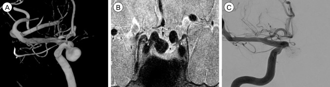
Fig. 1 (A) Three-dimensional (3D) DSA reconstruction shows a 7.9 mm aneurysm with medial projection at the right paraclinoid ICA. (B) PD MR coronal imaging shows the aneurysm (arrow head), distal dural ring/diaphragm (open arrow head), and cerebrospinal fluid space (star). (C) Successful coiling of the right paraclinoid aneurysm. DSA = digital subtraction angiography; ICA = internal carotid artery; PD MR = proton density magnetic resonance.
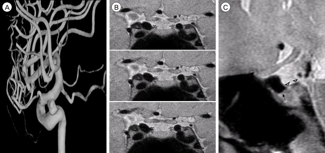
Fig. 2 (A) Three-dimensional (3D) DSA reconstruction shows a 5.0 mm aneurysm with an inferior/posterior projection at the right paraclinoid ICA. (B) PD MR coronal imaging cannot discriminate between the distal dural ring (open arrow head) and aneurysm (arrow). (C) PD MR multiplanar reconstruction shows the distal dural ring (open arrow head) and a transdural aneurysm (arrow). DSA = digital subtraction angiography; ICA = internal carotid artery; PD MR = proton density magnetic resonance.
Cited by 1 articles
-
Added Predictive Values of Proton Density Magnetic Resonance Imaging on Posterior Communicating Artery Aneurysms and Surrounding Soft Tissues with Simple Classification
Sun Yoon, Min Jeoung Kim, Hyun Jin Han, Keun Young Park, Joonho Chung, Yong Bae Kim
J Korean Neurosurg Soc. 2023;66(4):418-425. doi: 10.3340/jkns.2022.0259.
Reference
-
1. Bushong SC, Clarke G. Magnetic resonance imaging: physical and biological principles. St. Louis, MO: Mosby;2013. p. 64–66.2. Hashimoto K, Nozaki K, Hashimoto N. Optic strut as a radiographic landmark in evaluating neck location of a paraclinoid aneurysm. Neurosurgery. 2006; 10. 59(4):880–895. discussion 896-7PMID: 17038952.
Article3. Hirai T, Kai Y, Morioka M, Yano S, Kitajima M, Fukuoka H, et al. Differentiation between paraclinoid and cavernous sinus aneurysms with contrast-enhanced 3D constructive interference in steady-state MR imaging. AJNR Am J Neuroradiol. 2008; 1. 29(1):130–133. PMID: 17974619.4. Horowitz AL. MRI Physics for Radiologists: A Visual Approach. New York: Springer-Verlag;1992. p. 49–50.5. Jeon HJ, Lee JW, Kim SY, Park KY, Huh SK. Morphological parameters related to ruptured aneurysm in the patient with multiple cerebral aneurysms (clinical investigation). Neurol Res. 2014; 12. 36(12):1056–1062. PMID: 24852695.
Article6. Jeon JS, Ahn JH, Huh W, Son YJ, Bang JS, Kang HS, et al. A retrospective analysis on the natural history of incidental small paraclinoid unruptured aneurysm. J Neurol Neurosurg Psychiatry. 2014; 3. 85(3):289–294. PMID: 23781005.
Article7. Lee N, Jung JY, Huh SK, Kim DJ, Kim DI, Kim J. Distinction between Intradural and Extradural Aneurysms Involving the Paraclinoid Internal Carotid Artery with T2-Weighted Three-Dimensional Fast Spin-Echo Magnetic Resonance Imaging. J Korean Neurosurg Soc. 2010; 6. 47(6):437–441. PMID: 20617089.
Article8. Murayama Y, Sakurama K, Satoh K, Nagahiro S. Identification of the carotid artery dural ring by using three-dimensional computerized tomography angiography. J Neurosurg. 2001; 9. 95(3):533–536. PMID: 11565882.
Article9. Thines L, Lee SK, Dehdashti AR, Agid R, Willinsky RA, Wallace CM, et al. Direct imaging of the distal dural ring and paraclinoid internal carotid artery aneurysms with high-resolution T2 turbo-spin echo technique at 3-T magnetic resonance imaging. Neurosurgery. 2009; 6. 64(6):1059–1064. discussion 1064PMID: 19487884.
Article10. Watanabe Y, Nakazawa T, Yamada N, Higashi M, Hishikawa T, Miyamoto S, et al. Identification of the distal dural ring with use of fusion images with 3D-MR cisternography and MR angiography: application to paraclinoid aneurysms. AJNR Am J Neuroradiol. 2009; 4. 30(4):845–850. PMID: 19147723.
Article
- Full Text Links
-
- Actions
-
Cited
- CITED
-
- Close
- Share
-
- Similar articles
-
- Analysis of Clinical Outcome and Complications After Microsurgical Clipping of Unruptured Paraclinoid Aneurysms
- Progressive Visual Loss after Endovascular Coiling Treatment of a Large Paraclinoid Aneurysm
- Unruptured Paraclinoid Carotid Aneurysms Occur More Frequently in Younger Ages
- Frequency and Characteristics of Paraclinoid Aneurysm in Ruptured Cerebral Aneurysms
- Distinction between Intradural and Extradural Aneurysms Involving the Paraclinoid Internal Carotid Artery with T2-Weighted Three-Dimensional Fast Spin-Echo Magnetic Resonance Imaging
