J Korean Soc Spine Surg.
2015 Sep;22(3):109-113. 10.4184/jkss.2015.22.3.109.
The Use of Vessel Loop Shoelace Technique for Closure of Wound Dehiscence Caused by Dural Tears Associated with Distractive Flexion Injury of Cervical Spine
- Affiliations
-
- 1Department of Orthopaedic Surgery, School of Medicine, Chosun University, Gwangju, Korea. wwwpibak@daum.net
- KMID: 2068929
- DOI: http://doi.org/10.4184/jkss.2015.22.3.109
Abstract
- STUDY DESIGN: A case report.
OBJECTIVES
To report the use of the shoelace technique for treatment of wound dehiscence caused by dural tears. SUMMARY OF LITERATURE REVIEW: It is difficult to treat wound dehiscence caused by dural tears, as it can lead to infection, loss of soft tissue, and need for a long hospital stay.
MATERIALS AND METHODS
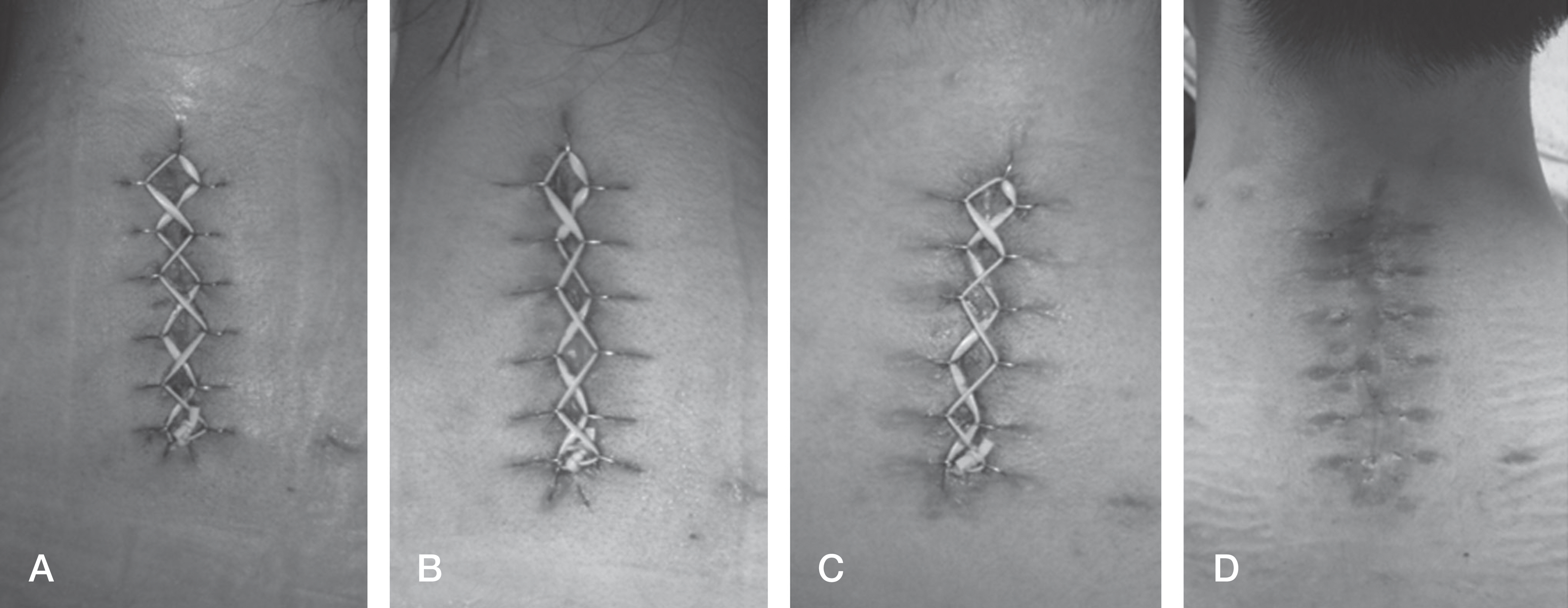
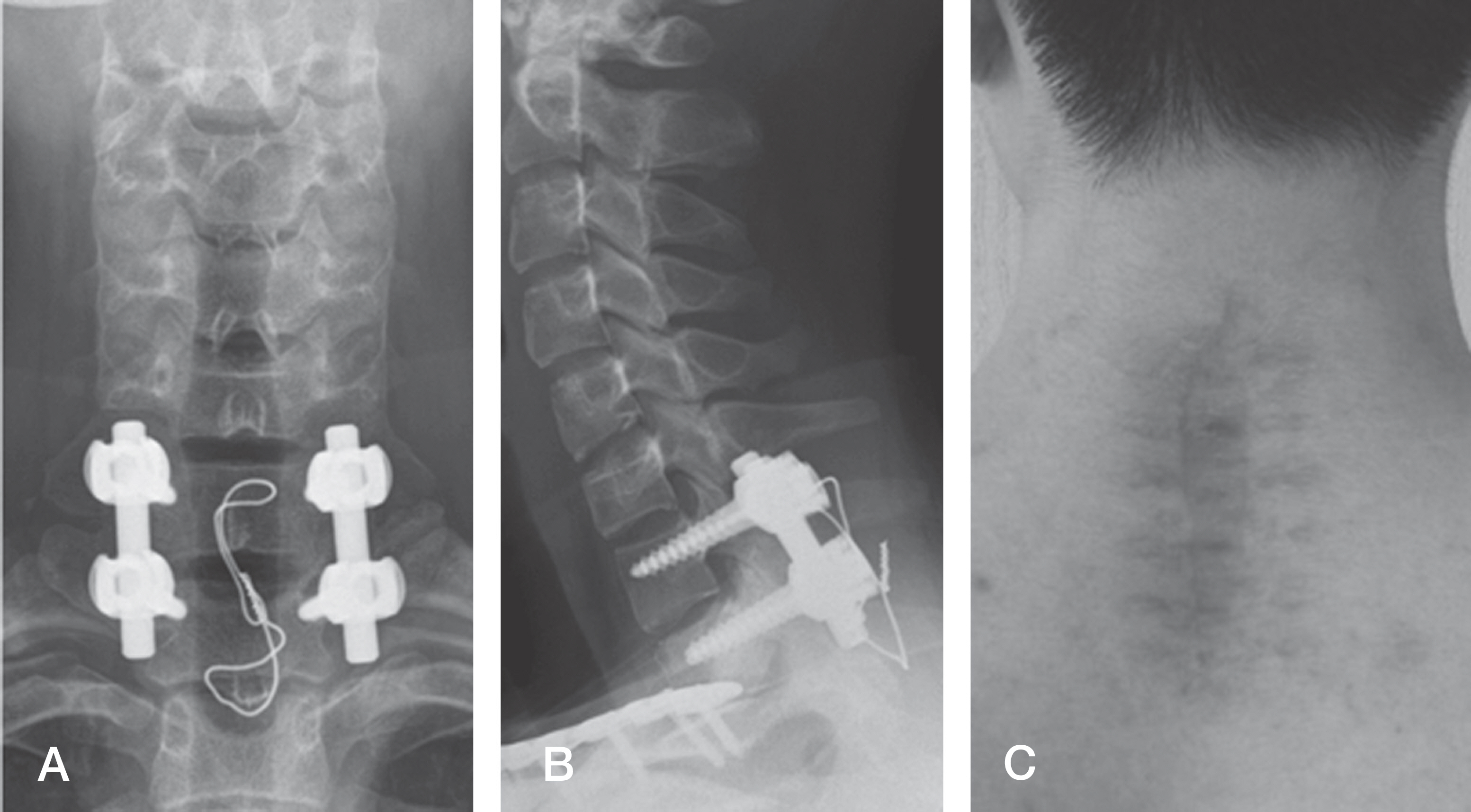
An 18-year-old male who had been injured in a traffic accident was diagnosed with bilateral facet dislocation of C7-T1, with no neurologic deficit. Clear secretion appeared during the operation, but it disappeared after posterior fusion. The wound began to open about 3 weeks after the operation. We used the vessel loop shoelace technique to suture the wound.
RESULTS
The patient had the stitches taken out in the outpatient clinic three weeks after suture. His wounds are healing without complication.
CONCLUSIONS
The vessel loop shoelace technique may be a useful treatment for wound dehiscence caused by dural tears.
MeSH Terms
Figure
Reference
-
1. Luszczyk MJ, Blaisdell GY, Wiater BP, et al. Traumatic dural tears: what do we know and are they a problem? Spine J. 2014; 14:49–56.
Article2. Khan MH, Rihn J, Steele G, et al. Postoperative management protocol for incidental dural tears during degenerative lumbar spine surgery: a review of 3,183 consecutive degenerative lumbar cases. Spine (Phila Pa 1976). 2006; 31:2609–13.3. Wang JC, Bohlman HH, Riew KD. Dural tears secondary to operations on the lumbar spine. Management and results after a two-year-minimum followup of eighty-eight patients. J Bone Joint Surg Am. 1998; 80:1728–32.
Article4. Fountas KN, Kapsalaki EZ, Johnston KW. Cerebrospinal fluid fistula secondary to dural tear in anterior cervical discectomy and fusion: case report. Spine (Phila Pa 1976). 2005; 30:E277–80.5. Cammisa FP, Jr., Girardi FP, Sangani PK, Parvataneni HK, 6. Hannallah D, Lee J, Khan M, Donaldson WF, Kang JD. Cerebrospinal fluid leaks following cervical spine surgery. J Bone Joint Surg Am. 2008; 90:1101–5.7. Asgari MM, Spinelli HM. The vessel loop shoelace technique for closure of fasciotomy wounds. Ann Plast Surg. 2000; 44:225–9.
Article8. Eid A, Elsoufy M. Shoelace wound closure for the management of fracture-related fasciotomy wounds. ISRN Orthop. 2012; 2012:528382.
Article9. Mahindra P, Bassi JL, Bassi D, et al. Shoe lace suture technique for management of lower limb diabetic amputations- a study of 24 cases. Pb Journal of Orthopaedics. 2011; 7:31–3.10. Fowler JR, Kleiner MT, Das R, Gaughan JP, Rehman S. Assisted closure of fasciotomy wounds: A descriptive series and caution in patients with vascular injury. Bone Joint Res. 2012; 1:31–5.
- Full Text Links
- Actions
-
Cited
- CITED
-
- Close
- Share
- Similar articles
-
- Combining Negative Pressure Wound Therapy with Shoelace Technique for Effective Closure of Soft Tissue Defect: A Case Series
- Injury of Posterior Ligament Complex with Cervical Spine Fracture
- Delayed Diagnosed Stage 1, 2 Distractive Flexion Injury of the Cervical Spine
- Treatment of Distractive Flexion Injury in Lower Cervical Spine using Anterior Cervical Fusion
- Clinical Outcomes of Fasciotomy for Acute Compartment Syndrome


