J Korean Soc Surg Hand.
2015 Sep;20(3):133-137. 10.12790/jkssh.2015.20.3.133.
The Importance of Preoperative Imaging Study on a Solitary Neurofibroma Originated from the Digital Nerve
- Affiliations
-
- 1Department of Plastic Surgery, Seoul St. Mary's Hospital, College of Medicine, The Catholic University of Korea, Seoul, Korea. nasuko@catholic.ac.kr
- KMID: 2068819
- DOI: http://doi.org/10.12790/jkssh.2015.20.3.133
Abstract
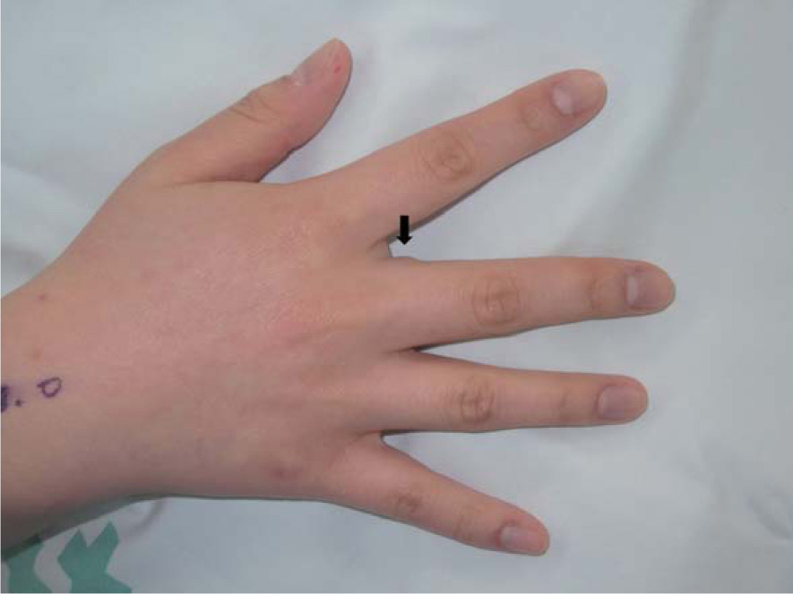
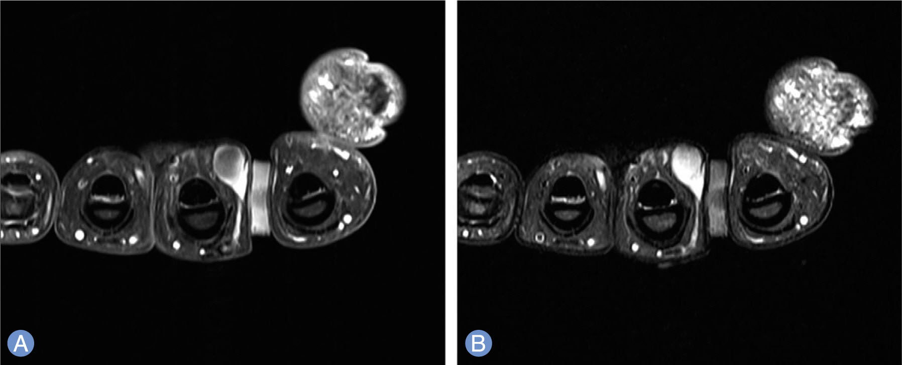
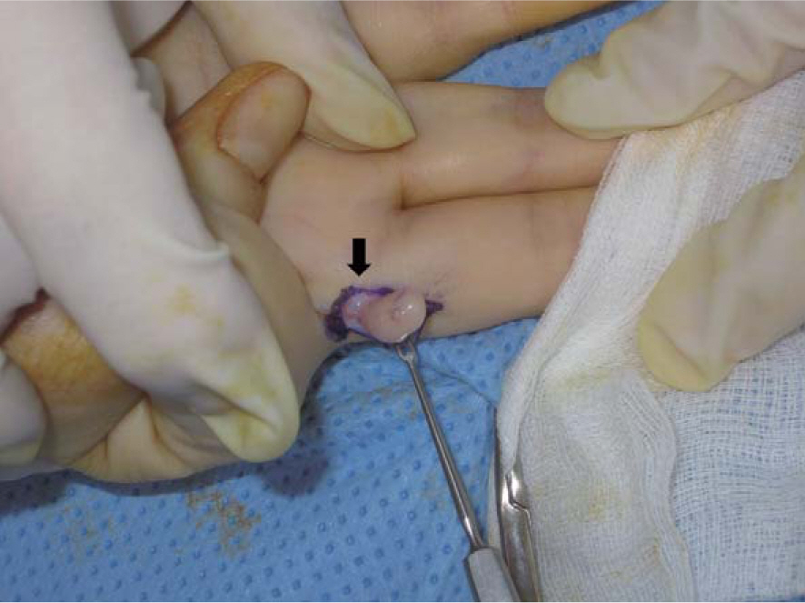
- This case is about a rare type of a solitary neurofibroma that originated from the digital nerve between the proximal phalanx of a finger and the web space, which was first misdiagnosed as giant cell tumor, ganglionic cyst, or fibroma originating from the tendon before radiologic studies were done. The preoperative magnetic resonance imaging (MRI) showed a non-enhanced well-circumscribed mass and the digital nerve was deviated to the volar-medial side due to the mass effect. Since neurofibroma is difficult to differentiate from others by physical examination, crucial information such as the connection between the mass and the nerve or the deviation of the digital nerve can be obtained by MRI findings. And it is important to plan the surgery safely from this information.
Keyword
MeSH Terms
Figure
Reference
-
References
1. Huajun J, Wei Q, Ming L, Chongyang F, Weiguo Z, Decheng L. Solitary subungual neurofibroma in the right first finger. Int J Dermatol. 2012; 51:335–8.
Article2. Seo BM, Lim JS, Jung SN, Yoo G, Byeon JH. Solitary subungual myxoid neurofibroma of the thumb: a case report. J Korean Soc Plast Reconstr Surg. 2011; 38:398–400.3. Gerber PA, Antal AS, Neumann NJ, et al. Neurofibromatosis. Eur J Med Res. 2009; 14:102–5.
Article4. Dangoisse C, Andre J, De Dobbeleer G, Van Geertruyden J. Solitary subungual neurofibroma. Br J Dermatol. 2000; 143:1116–7.
Article5. Baran R, Haneke E. Subungual myxoid neurofibroma on the thumb. Acta Derm Venereol. 2001; 81:210–1.6. Bhushan M, Telfer NR, Chalmers RJ. Subungual neurofibroma: an unusual cause of nail dystrophy. Br J Dermatol. 1999; 140:777–8.7. Niizuma K, Iijima KN. Solitary neurofibroma: a case of subungual neurofibroma on the right third finger. Arch Dermatol Res. 1991; 283:13–5.
Article8. Chong AK, Tan DM. Diagnostic imaging of the hand and wrist. Neligan P, editor. Plastic surgery. New York: Elsevier Saunders;2012; 78–83.9. Wang Y, Tang J, Luo Y. The value of sonography in diagnosing giant cell tumors of the tendon sheath. J Ultrasound Med. 2007; 26:1333–40.
Article10. Purohit S, Pardiwala DN. Imaging of giant cell tumor of bone. Indian J Orthop. 2007; 41:91–6.
Article
- Full Text Links
- Actions
-
Cited
- CITED
-
- Close
- Share
- Similar articles
-
- Solitary Plexiform Neurofibroma on the Median Nerve: A Case Report
- Solitary Neurofibroma in the Orbit
- Solitary neurofibroma of the sciatic nerve which was initially misdiagnosed as herniated nucleus pulposus: A case report
- A Giant Retroperitoneal Neurofibroma
- A Case of Solitary Neurofibroma on the Finger with Nail Deformity


