Frenectomy for improvement of a problematic conventional maxillary complete denture in an elderly patient: a case report
- Affiliations
-
- 1Dental Biomaterials Research and Development Chair, College of Dentistry, King Saud University College of Dentistry, Riyadh, Saudi Arabia. yaljabbari@ksu.edu.sa
- 2Department of Prosthetic Dental Sciences, College of Dentistry, King Saud University College of Dentistry, Riyadh, Saudi Arabia.
- KMID: 2054759
- DOI: http://doi.org/10.4047/jap.2011.3.4.236
Abstract
- Maxillary labial and buccal frena are considered as normal anatomic structures in the oral cavity. However, they may exist intraorally as a thick broad fibrous attachment and/or become located near the crest of the residual ridge, thus interfering with proper denture border extension resulting in inferior denture stability, retention and overall patient satisfaction. This case report highlights the importance of clinical examination and treatment planning which may mandate preprosthetic surgery prior to fabrication of a new conventional complete denture. Adequate patient satisfaction with conventional complete dentures can be significantly increased after frenectomy.
Keyword
MeSH Terms
Figure
-
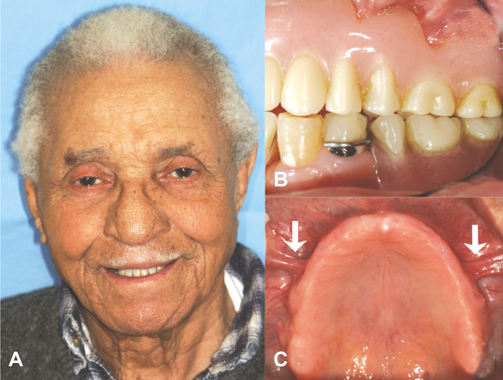
Fig. 1 Pre-treatment views. A: Extra-oral view with the existing prosthesis, B: Intra-oral left lateral view of the complete denture, C: Occlusal view of the maxilla showing low and thick buccal frena (arrows).
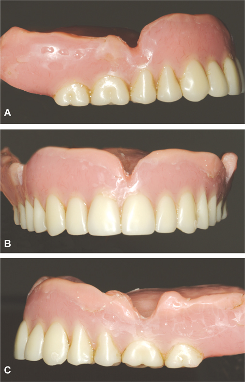
Fig. 2 Existing patient's maxillary denture after chair-side hard relining. A: Right lateral view, B: Frontal view, C: Left lateral view. Note the significant relief areas (labial and buccal notches) that were required to accommodate the low and thick frena.
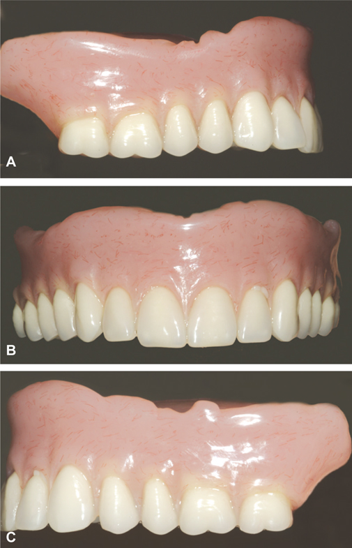
Fig. 3 Newly fabricated maxillary denture after frenectomy. A: Right lateral view, B: Frontal view, C: Left lateral view. Note the improved borders extension of the denture compared to what is shown in Fig. 2.
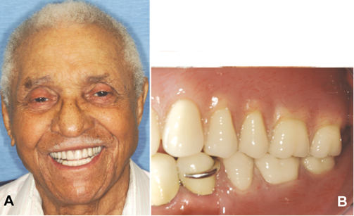
Fig. 4 Post-treatment views. A: Extra-oral view with the newly fabricated prosthesis, B: Intra-oral left lateral view of the complete denture.
Reference
-
1. Douglass CW, Shih A, Ostry L. Will there be a need for complete dentures in the United States in 2020? J Prosthet Dent. 2002. 87:5–8.2. Zarb GA, Carlsson GE, Bolender CL. Boucher's prosthodontic treatment for edentulous patients. 1997. 11th ed. St. Louis: Mosby;46–47.3. Carlsson GE, Omar R. The future of complete dentures in oral rehabilitation. A critical review. J Oral Rehabil. 2010. 37:143–156.4. Christensen GJ. Treatment of the edentulous mandible. J Am Dent Assoc. 2001. 132:231–233.5. van Waas MA. The influence of clinical variables on patients' satisfaction with complete dentures. J Prosthet Dent. 1990. 63:307–310.6. Hillerup S. Preprosthetic surgery in the elderly. J Prosthet Dent. 1994. 72:551–558.7. Axinn S, Brasher WJ. Frenectomy plus free graft. J Prosthet Dent. 1983. 50:16–19.8. Costello BJ, Betts NJ, Barber HD, Fonseca RJ. Preprosthetic surgery for the edentulous patients. Dent Clin North Am. 1996. 40:19–38.9. Driscoll CF, Masri RM. Single maxillary complete denture. Dent Clin North Am. 2004. 48:567–583.
- Full Text Links
-
- Actions
-
Cited
- CITED
-
- Close
- Share
-
- Similar articles
-
- Full mouth rehabilitation of an edentulous patient using maxillary complete denture and mandibular implant supported fixed prostheses: a case report
- Phonetic improvement by adjusting the shape of the anterior palate of the maxillary complete denture: a case report
- Comparison of conventional and suction-effective complete denture in a fully edentulous patient: a case report
- The Implant Retained Overdenture by Locator Attachments on the Edentulous Mandible: A Case Report
- Maxillary complete denture fabrication cases with posterior palatal seal considering palatal form and tissue displacement
