Hip Pelvis.
2014 Mar;26(1):36-40. 10.5371/hp.2014.26.1.36.
Arthroscopic Fragment Excision of Pipkin Type I Displaced Femoral Head Fracture: A Case Report
- Affiliations
-
- 1Department of Orthopaedic Surgery, St. Carollo Hospital, Suncheon, Korea. wctoilets@hanmail.net
- KMID: 2054160
- DOI: http://doi.org/10.5371/hp.2014.26.1.36
Abstract
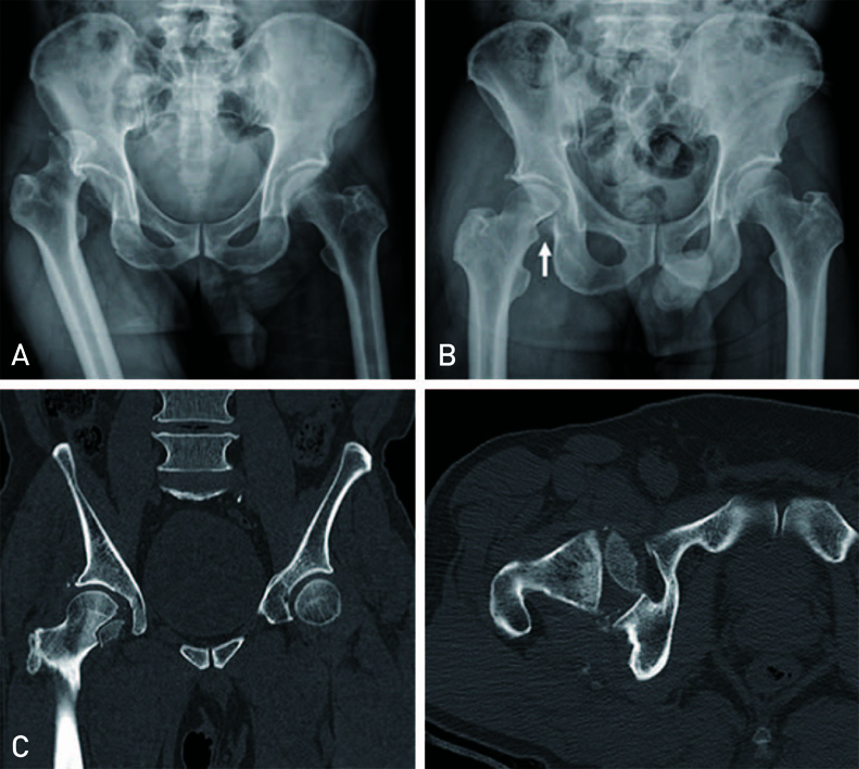
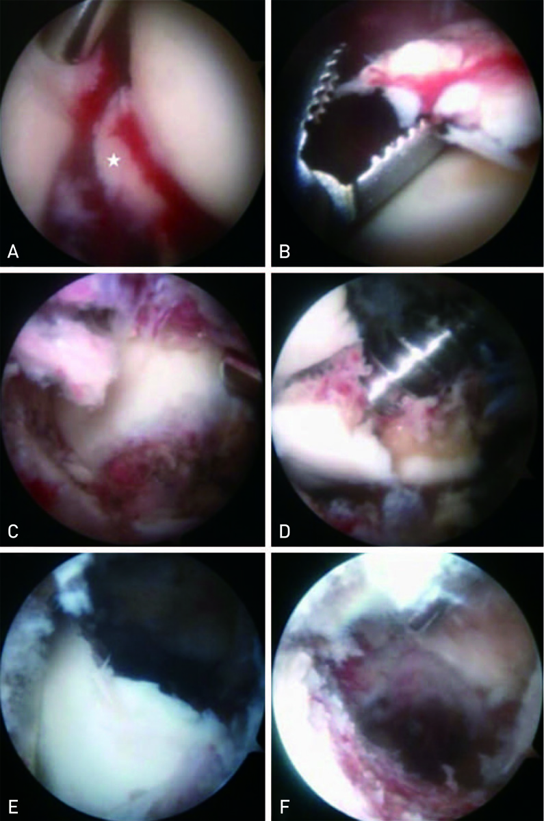
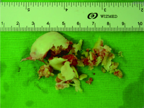
- There has been a variety of options for treatment of femoral head fracture with hip dislocation according to the Pipkin classification. Pipkin type I fractures with minimal displacement have been treated conservatively. However, in cases where the fracture was displaced or reduced incongruently, it has been treated by open fragment excision or fixation after reduction. In our case, the patient was a 62-year-old man who sustained a displaced fracture of Pipkin type I. We achieved a satisfactory outcome by arthroscopic excision of a displaced bony fragment and small bony fragments that could not be confirmed by pre-operative imaging study. Therefore, we report on the case with a review of the literature.
Figure
Reference
-
1. Sahin V, Karakaş ES, Aksu S, Atlihan D, Turk CY, Halici M. Traumatic dislocation and fracture-dislocation of the hip: a long-term follow-up study. J Trauma. 2003; 54:520–529.
Article2. Lansford T, Munns SW. Arthroscopic treatment of Pipkin type I femoral head fractures: a report of 2 cases. J Orthop Trauma. 2012; 26:e94–e96.3. Park MS, Her IS, Cho HM, Chung YY. Internal fixation of femoral head fractures (Pipkin I) using hip arthroscopy. Knee Surg Sports Traumatol Arthrosc. Published online January 9, 2014; doi:10.1007/s00167-013-2821-4.
Article4. Matsuda DK. A rare fracture, an even rarer treatment: the arthroscopic reduction and internal fixation of an isolated femoral head fracture. Arthroscopy. 2009; 25:408–412.
Article5. Mullis BH, Dahners LE. Hip arthroscopy to remove loose bodies after traumatic dislocation. J Orthop Trauma. 2006; 20:22–26.
Article6. Asghar FA, Karunakar MA. Femoral head fractures: diagnosis, management, and complications. Orthop Clin North Am. 2004; 35:463–472.
Article7. Stannard JP, Harris HW, Volgas DA, Alonso JE. Functional outcome of patients with femoral head fractures associated with hip dislocations. Clin Orthop Relat Res. 2000; (377):44–56.
Article8. Chen ZW, Lin B, Zhai WL, et al. Conservative versus surgical management of Pipkin type I fractures associated with posterior dislocation of the hip: a randomised controlled trial. Int Orthop. 2011; 35:1077–1081.
Article9. Keene GS, Villar RN. Arthroscopic loose body retrieval following traumatic hip dislocation. Injury. 1994; 25:507–510.
Article10. Svoboda SJ, Williams DM, Murphy KP. Hip arthroscopy for osteochondral loose body removal after a posterior hip dislocation. Arthroscopy. 2003; 19:777–781.
Article
- Full Text Links
- Actions
-
Cited
- CITED
-
- Close
- Share
- Similar articles
-
- Posterior Fracture Dislocation of the Hip with Fracture of the Femoral Head
- Fixation of Pipkin Fractures with Acutrak Screws: A Report of Three Cases
- Result of Surgical Treatment for the Femoral Head Fracture
- Femoral Head Fractures
- Relevance of the Watson-Jones anterolateral approach in the management of Pipkin type II fracture-dislocation: a case report and literature review


