Fatal Small Bowel Bleeding with very Low Risk Gastrointestinal Stromal Tumor in Jejunum
- Affiliations
-
- 1Department of Internal Medicine, Kyung Hee University Hospital at Gangdong, Kyung Hee University School of Medicine, Seoul, Korea. drcha@khu.ac.kr
- KMID: 2052784
- DOI: http://doi.org/10.12771/emj.2015.38.2.72
Abstract
- Gastrointestinal stromal tumor (GIST) is the most common mesenchymal neoplasm of the gastrointestinal (GI) tract. These tumors are frequently small, asymptomatic and found incidentally. GI bleeding is a common complication of these tumors, but small sized, very low risk GIST rarely complicated with fatal bleeding. In this report, we describe a 42-year-old woman with a jejunal GIST accompanied by severe GI bleeding. She presented with melena and an angiographic embolization was performed for a jejunal mass with bleeding. However, rebleeding was suspected after an angiographic embolization and an emergent segmental resection for the bleeding mass was performed. She was finally diagnosed as a 1.8 cm sized very low risk GIST in jejunum. In conclusion, physician should consider that even very low risk GIST can be the cause of GI bleeding when there is severe bleeding.
Keyword
Figure
-
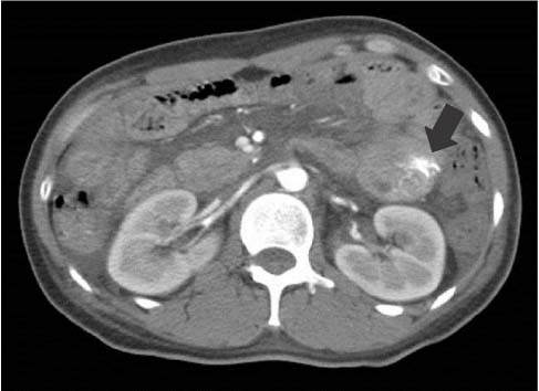
Fig. 1 Abdominal computed tomography finding. It shows extravasation of contrast agent with 1.5 cm sized filling detect (black arrow). It allows assessment of active bleeding in proximal jejunal loop with small mass.
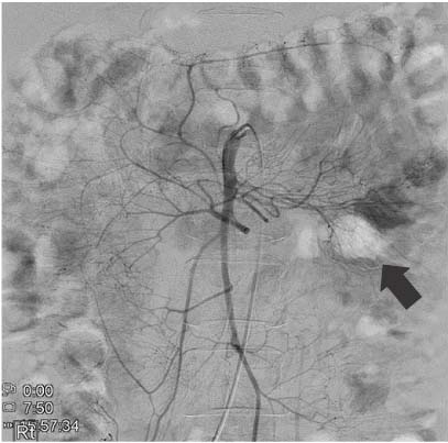
Fig. 2 Superior mesenteric artery angiography. Active extravasation is noted from the 1st jejunal branch (black arrow).
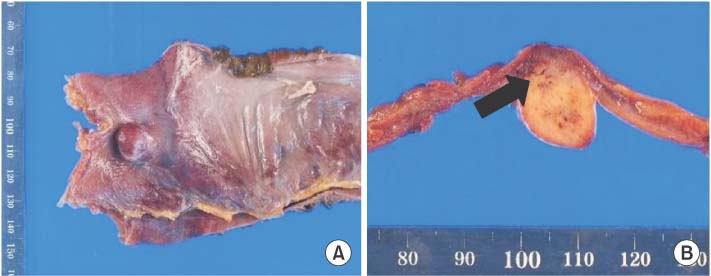
Fig. 3 Gross findings of resected specimen. (A) It shows a 1.8×1.5×1.5 cm sized, encapsulated lobulating mass with hemorrhage. (B) The cut surface shows a gray-white solid tumor with multifocal hemorrhages (black arrow).

Fig. 4 Microscopic findings of resected specimen. (A) It reveals spindle shaped cells arranged in fascicular pattern (H&E, ×400). (B) In immunohistochemical staining, the tumor cells are positive for c-kit (c-kit stain, ×200).
Reference
-
1. Pidhorecky I, Cheney RT, Kraybill WG, Gibbs JF. Gastrointestinal stromal tumors: current diagnosis, biologic behavior, and management. Ann Surg Oncol. 2000; 7:705–712.2. Joensuu H. Gastrointestinal stromal tumor (GIST). Ann Oncol. 2006; 17:Suppl 10. x280–x286.3. Singhal T, Doddi S, Leake T, Parsi S, Hussain A, Chandra A, et al. Upper gastrointestinal bleeding due to gastric stromal tumour: a case report. Cases J. 2010; 3:58.4. Daldoul S, Moussi A, Triki W, Baraket RB, Zaouche A. Jejunal GIST causing acute massive gastrointestinal bleeding: role of multidetector row helical CT in the preoperative diagnosis and management. Arab J Gastroenterol. 2012; 13:153–157.5. Kramer K, Siech M, Strater J, Aschoff AJ, Henne-Bruns D. GI hemorrhage with fulminant shock induced by jejunal gastrointestinal stromal tumor (GIST) coincident with duodenal neuroendocrine carcinoma (NET) + neurofibromatosis (NF): case report and review of the literature. Z Gastroenterol. 2005; 43:281–288.6. Nilsson B, Bümming P, Meis-Kindblom JM, Oden A, Dortok A, Gustavsson B, et al. Gastrointestinal stromal tumors: the incidence, prevalence, clinical course, and prognostication in the preimatinib mesylate era: a population-based study in western Sweden. Cancer. 2005; 103:821–829.7. Jhu IK, Joo YE, Park GS, Park MH, Park SU, Lee NH, et al. A case of duodenal gastrointestinal stromal tumor presenting with gastrointestinal bleeding. Korean J Gastrointest Endosc. 2005; 31:121–125.8. Judson I. Gastrointestinal stromal tumours (GIST): biology and treatment. Ann Oncol. 2002; 13:Suppl 4. 287–289.9. Choi H, Choi KY, Eun CS, Jang HJ, Park DI, Chang DK, et al. Korean experience with double balloon endoscopy: Korean Association for the Study of Intestinal Diseases multi-center study. Gastrointest Endosc. 2007; 66:3 Suppl. S22–S25.10. Yoon W, Jeong YY, Shin SS, Lim HS, Song SG, Jang NG, et al. Acute massive gastrointestinal bleeding: detection and localization with arterial phase multi-detector row helical CT. Radiology. 2006; 239:160–167.11. Tew K, Davies RP, Jadun CK, Kew J. MDCT of acute lower gastrointestinal bleeding. AJR Am J Roentgenol. 2004; 182:427–430.12. Bensimhon D, Soyer P, Boudiaf M, Fargeaudou Y, Nemeth J, Pocard M, et al. Imaging of gastrointestinal stromal tumors. J Radiol. 2009; 90:469–480.13. Sass DA, Chopra KB, Finkelstein SD, Schauer PR. Jejunal gastrointestinal stromal tumor: a cause of obscure gastrointestinal bleeding. Arch Pathol Lab Med. 2004; 128:214–217.14. Majdoub Hassani KI, Zahid FZ, Ousadden A, Mazaz K, Taleb KA. Gastrointestinal stromal tumors and shock. J Emerg Trauma Shock. 2009; 2:199–202.15. Fletcher CD, Berman JJ, Corless C, Gorstein F, Lasota J, Longley BJ, et al. Diagnosis of gastrointestinal stromal tumors: a consensus approach. Hum Pathol. 2002; 33:459–465.
- Full Text Links
-
- Actions
-
Cited
- CITED
-
- Close
- Share
-
- Similar articles
-
- A Case of Bleeding from a Jejunal Gastrointestinal Stromal Tumor Diagnosed by Double Balloon Enteroscopy
- Multiple Gastrointestinal Stromal Tumors of the Small Intestine
- A Case of Massive Bleeding from Jejunal Stromal Tumor Diagnosed by Intraoperative Enteroscopy: A Case of Jejunal Stromal Tumor Bleeding
- Two Cases of Jejunal Gastrointestinal Stromal Tumor Diagnosed by Capsule Endoscope
- Primary Gastrointestinal Stromal Tumor Accompanied with Gastrointestinal Bleeding
