Intramural Gastric Abscess Caused by a Toothpick Presenting as a Subepithelial Tumor
- Affiliations
-
- 1Division of Gastrointestinology, Department of Internal Medicine, Presbyterian Medical Center, Jeonju, Korea. jeja-1004@hanmail.net
- KMID: 2049022
- DOI: http://doi.org/10.5946/ce.2014.47.3.254
Abstract
- In the present report, we describe an unusual case of an intramural gastric abscess caused by a foreign body, detected in the form of a subepithelial tumor. A 64-year-old woman was referred to our gastroenterology clinic for further evaluation of a gastric subepithelial tumor. The patient presented with a 1-month history of sustained dull epigastric pain. Esophagogastroduodenoscopy revealed an ill-demarcated, round, smooth, protruding lesion with a small central erosion on the great curvature of the proximal antrum. Endoscopic ultrasonography indicated the presence of an ovoid, heterogeneous, hypoechoic lesion with small echogenic foci located in the submucosa and muscularis propria layers. An abdominal computed tomography scan showed focal gastric wall thickening and regional lymph node enlargement. Endoscopic submucosal dissection was performed for definite diagnosis and management. Thus, we detected a toothpick and removed it using grasping forceps. The final diagnosis was an intramural gastric abscess caused by a toothpick.
Keyword
MeSH Terms
Figure
-
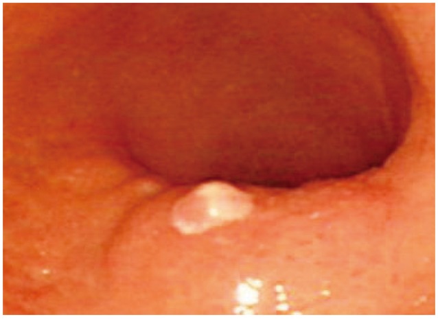
Fig. 1 Endoscopic findings. An ill-demarcated, round, smooth, protruding lesion with a small central erosion in the great curvature of the proximal antrum.
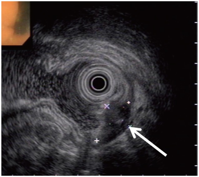
Fig. 2 Endoscopic ultrasonography findings. An ovoid, heterogeneous, hypoechoic mass with small echogenic foci located in the submucosa and muscularis propria layers (arrow).
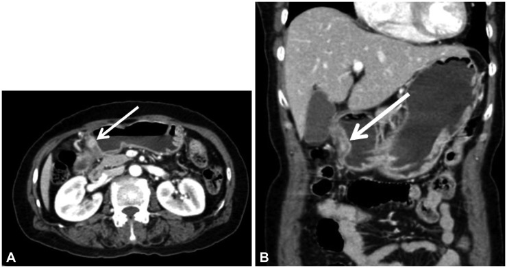
Fig. 3 Abdominal computed tomography scan images. (A) Horizontal view image and (B) sagittal view image show focal gastric wall thickening with heterogeneous enhancement (arrow) and regional lymph node enlargement. Abdominal perforation or major vessel injury was not evident.
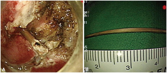
Fig. 4 Endoscopic submucosal dissection findings. (A) A toothpick that penetrated the antral wall of the stomach. (B) The 3-cm long toothpick, after removal.
Reference
-
1. Meltzer SJ, Goldberg MD, Meltzer RM, Claps F. Appendiceal obstruction by a toothpick removed at colonoscopy. Am J Gastroenterol. 1986; 81:1107–1108. PMID: 3776965.2. Abel RM, Fischer JE, Hendren WH. Penetration of the alimentary tract by a foreign body with migration to the liver. Arch Surg. 1971; 102:227–228. PMID: 5544643.
Article3. Budnick LD. Toothpick-related injuries in the United States, 1979 through 1982. JAMA. 1984; 252:796–797. PMID: 6748180.
Article4. Guber MD, Suarez CA, Greve J. Toothpick perforation of the intestine diagnosed by a small bowel series. Am J Gastroenterol. 1996; 91:789–791. PMID: 8677952.5. Porcu A, Dessanti A, Feo CF, Dettori G. Asymptomatic gastric perforation by a toothpick. A case report. Dig Surg. 1999; 16:437–438. PMID: 10567809.6. Meyns BP, Faveere BC, Van de, Dotremont G, Daenen WJ. Constrictive pericarditis due to ingestion of a toothpick. Ann Thorac Surg. 1994; 57:489–490. PMID: 8311624.
Article7. Matsubara M, Hirasaki S, Suzuki S. Gastric penetration by an ingested toothpick successfully managed with computed tomography and endoscopy. Intern Med. 2007; 46:971–974. PMID: 17603235.
Article8. Singh AC, Gurney M. Toothpick penetration of stomach. Gastrointest Endosc. 2003; 57:239. PMID: 12556792.
Article9. Rioux M, Langis P. Sonographic detection of clinically unsuspected swallowed toothpicks and their gastrointestinal complications. J Clin Ultrasound. 1994; 22:483–490. PMID: 7814653.
Article10. Hwang JH, Kimmey MB. The incidental upper gastrointestinal subepithelial mass. Gastroenterology. 2004; 126:301–307. PMID: 14699508.
Article11. Park JH, Kim HW, Park WI, et al. A case of gastric wall abscess associated with gastritis cystica profunda. Korean J Gastrointest Endosc. 2004; 29:509–513.
- Full Text Links
-
- Actions
-
Cited
- CITED
-
- Close
- Share
-
- Similar articles
-
- A Case of Liver Abscess Caused by Toothpick Penetrating Gastric Wall
- Diagnosis of Gastric Subepithelial Tumor: Focusing on Endoscopic Ultrasonography
- A Case of Intramural Gastric Wall Abscess, a Rare Disease Successfully Treated with Endoscopic Incision and Drainage
- A Case of Liver Abscess Associated with Duodenal Perforation by a Toothpick
- Gastric Wall Abscess Caused by a Fish Bone and Treated with Endoscopic Management
