Ann Lab Med.
2015 Jul;35(4):445-448. 10.3343/alm.2015.35.4.445.
Evaluation of Modified Formalin-Ether Concentration Method Using Para Tube in Clinical Settings
- Affiliations
-
- 1Department of Laboratory Medicine, Chonnam National University Hospital, Gwangju, Korea. dwryang@chonnam.ac.kr
- 2Department of Parasitology, College of Medicine, Seonam University, Namwon, Korea.
- KMID: 2045868
- DOI: http://doi.org/10.3343/alm.2015.35.4.445
Abstract
- Conventional formalin-ether concentration method is a gold standard for the diagnosis of parasite infection. However, it may be time-consuming and laborious. We aimed to reveal the clinical usefulness of a modified formalin-ether concentration method using the Para Tube (KS Corporation, Korea) compared with the conventional method. A total of 117 fresh, unpreserved fecal samples composed to 90 negative controls and 27 positive controls with ova of Diphyllobothrium latum/D. nihonkaiense, ova of Clonorchis sinensis and cysts of Giardia lamblia were used in this study. Both methods showed comparable correct identification rate (87.2% for the Para Tube vs. 86.3% for the conventional method).When five samples were examined at once, the Para Tube method reduced the procedure time compared with the conventional method (19 min 58 sec vs. 23 min 18 sec, P=0.0286). We concluded that the modified formalin-ether concentration method using the Para Tube is a rapid, simple, and reliable fecal concentration method for clinical use.
Keyword
Figure
-
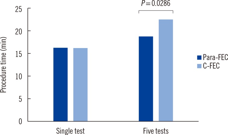
Fig. 1 Comparison of procedure time by the modified formalin-ether concentration using the Para Tube (Para-FEC) and the conventional formalin-ether concentration (C-FEC) methods, according to the increasing number of tests simultaneously. While the mean time for a single test was comparable in the two methods, the procedure time using the Para Tube was reduced compared with that required by the conventional method, when five samples were tested simultaneously.
Cited by 1 articles
-
Utility of an Automatic Vision-Based Examination System (AVE-562) for the Detection of
Clonorchis sinensis Eggs in Stool
Yu Jeong Lee, Eun Jeong Won, Young-Chang Cho, Soo Hyun Kim, Myung Geun Shin, Jong Hee Shin
Ann Lab Med. 2021;41(2):221-224. doi: 10.3343/alm.2021.41.2.221.
Reference
-
1. Stepek G, Buttle DJ, Duce IR, Behnke JM. Human gastrointestinal nematode infections: are new control methods required? Int J Exp Pathol. 2006; 87:325–341. PMID: 16965561.
Article2. Kim TS, Cho SH, Huh S, Kong Y, Sohn WM, Hwang SS, et al. A nationwide survey on the prevalence of intestinal parasitic infections in the Republic of Korea, 2004. Korean J Parasitol. 2009; 47:37–47. PMID: 19290090.
Article3. Barda BD, Rinaldi L, Ianniello D, Zepherine H, Salvo F, Sadutshang T, et al. Mini-FLOTAC, an innovative direct diagnostic technique for intestinal parasitic infections: experience from the field. PLoS Negl Trop Dis. 2013; 7:e2344. PMID: 23936577.
Article4. Smith JW, Bartlett MS. Diagnostic parasitology: introduction and methods. In : Lenette EH, Balows A, Hausler WJ, Shadomy HJ, editors. Manual of clinical microbiology. 4th ed. Washington, D.C.: American Society for Microbiology;1985. p. 595–611.
- Full Text Links
-
- Actions
-
Cited
- CITED
-
- Close
- Share
-
- Similar articles
-
- Modified Formalin-Ether Concentration Technique for Diagnosis of Human Strongyloidiasis
- Comparison of detection rate of some trematodes' eggs by combined technique of formalin-ether sedimentation and zinc sulfate flotation
- Endotracheal Tube Obstruction due to Mucous Crust during Inhalation Anesthesia
- Assessment of Modified Chromosome Analysis Method Using Culture Tube for Peripheral Blood
- An evaluation of cellophane thick smear technique for mass stool examination
