A Pedunculated Left Ventricular Thrombus in a Women with Peripartum Cardiomyopathy: Evaluation by Three Dimensional Echocardiography
- Affiliations
-
- 1Department of Cardiology, King Georges' Medical University, Lucknow, India. raj_iv_infusion@yahoo.co.in
- KMID: 2045430
- DOI: http://doi.org/10.4250/jcu.2014.22.3.139
Abstract
- Peripartum cardiomyopathy is a cardiac condition characterized by development of heart failure during the last month of pregnancy or during the first five months of post partum period without any other identifiable cause of heart failure. The hypercoagulable state in the pregnancy along with left ventricular (LV) systolic dysfunction predisposes the patient to thromboembolic complications like intraventricular thrombi. We report a case of a 30-year-old female with peripartum cardiomyopathy along with a highly mobile mass in the LV cavity on two dimensional echocardiography. Three dimensional transthoracic echocardiography clearly showed the pedicle of the mass attached to the interventricular septum along with internal echolucent areas within the mass. Due to denial of the patient to undergo surgery, she was started on oral anticoagulation, with complete dissolution of the mass within one month.
Keyword
MeSH Terms
Figure
-
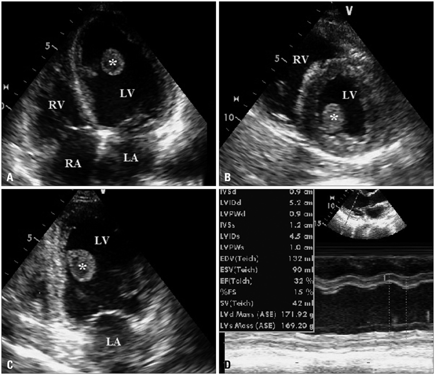
Fig. 1 Two dimensional transthoracic echocardiography. A large single pedunculated mass (indicated by *) can be seen in the LV cavity in apical four chamber (A), apical two chamber (C), and parasternal short axis (B) views. There is dilatation of all the four cardiac chambers with poor left ventricular ejection fraction of 32% (D). LA: left atrium, LV: left ventricle, RA: right atrium, RV: right ventricle.
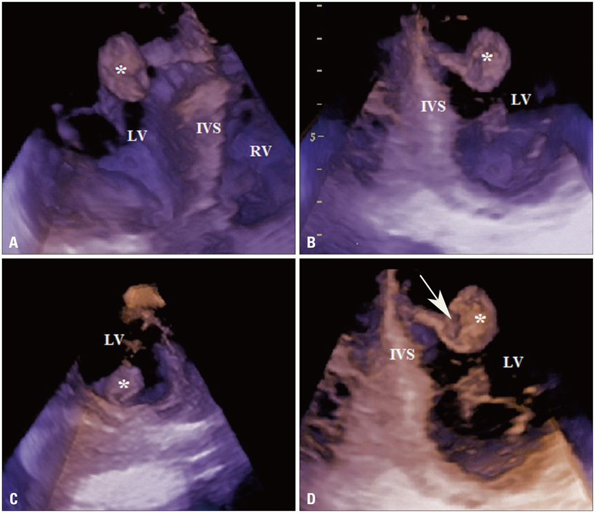
Fig. 2 Three dimensional (3D) transthoracic echocardiography. The mass within the LV cavity (indicated by *) can be viewed from different imaging planes (A-D) with the help of cropping the basic 3D data set. The site of attachment of the mass to the IVS via a pedicle, the irregular surface characteristics as well as the internal echo lucent areas within the mass (arrow in D) can be clearly delineated. IVS: interventricular septum, LV: left ventricle, RV: right ventricle.
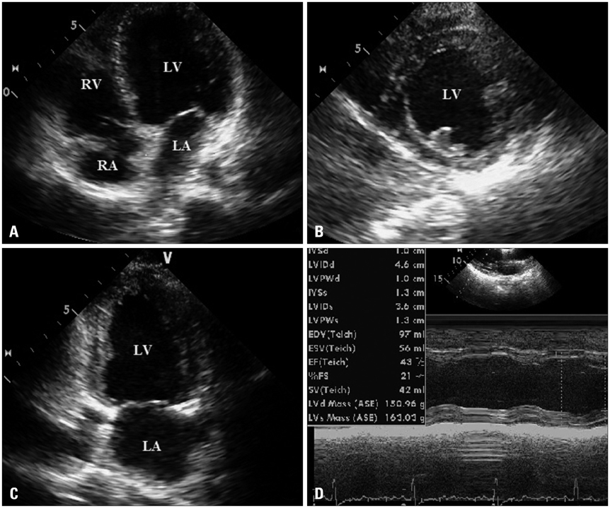
Fig. 3 Two dimensional transthoracic echocardiography. Follow-up echocardiography at 1 month after the onset of anticoagulation and heart failure therapy showed complete resolution of the intracardiac mass (A, B, and C) as well as improvement in the left ventricular ejection fraction (43%, D). LA: left atrium, LV: left ventricle, RA: right atrium, RV: right ventricle.
Reference
-
1. Demakis JG, Rahimtoola SH. Peripartum cardiomyopathy. Circulation. 1971; 44:964–968.
Article2. Cunningham FG, Pritchard JA, Hankins GD, Anderson PL, Lucas MJ, Armstrong KF. Peripartum heart failure: idiopathic cardiomyopathy or compounding cardiovascular events? Obstet Gynecol. 1986; 67:157–168.3. Bhattacharyya A, Basra SS, Sen P, Kar B. Peripartum cardiomyopathy: a review. Tex Heart Inst J. 2012; 39:8–16.4. Demakis JG, Rahimtoola SH, Sutton GC, Meadows WR, Szanto PB, Tobin JR, Gunnar RM. Natural course of peripartum cardiomyopathy. Circulation. 1971; 44:1053–1061.
Article5. Stratton JR, Lighty GW Jr, Pearlman AS, Ritchie JL. Detection of left ventricular thrombus by two-dimensional echocardiography: sensitivity, specificity, and causes of uncertainty. Circulation. 1982; 66:156–166.
Article6. Srichai MB, Junor C, Rodriguez LL, Stillman AE, Grimm RA, Lieber ML, Weaver JA, Smedira NG, White RD. Clinical, imaging, and pathological characteristics of left ventricular thrombus: a comparison of contrast-enhanced magnetic resonance imaging, transthoracic echocardiography, and transesophageal echocardiography with surgical or pathological validation. Am Heart J. 2006; 152:75–84.
Article7. Sinha A, Nanda NC, Khanna D, Dod HS, Vengala S, Mehmood F, Agrawal G, Upendram S. Morphological assessment of left ventricular thrombus by live three-dimensional transthoracic echocardiography. Echocardiography. 2004; 21:649–655.
Article8. Duncan K, Nanda NC, Foster WA, Mehmood F, Patel V, Singh A. Incremental value of live/real time three-dimensional transthoracic echocardiography in the assessment of left ventricular thrombi. Echocardiography. 2006; 23:68–72.
Article9. Nili M, Deviri E, Jortner R, Strasberg B, Levy MJ. Surgical removal of a mobile, pedunculated left ventricular thrombus: report of 4 cases. Ann Thorac Surg. 1988; 46:396–400.
Article10. Altuwaijri WA, Kirkpatrick ID, Jassal DS, Soni A. Vanishing left ventricular thrombus in a woman with peripartum cardiomyopathy: a case report. BMC Res Notes. 2012; 5:544.
Article
- Full Text Links
-
- Actions
-
Cited
- CITED
-
- Close
- Share
-
- Similar articles
-
- Surgical Removal of a Pedunculated Left Ventricular thrombus
- Apical Hypertrophic Cardiomyopathy with Apical Aneurysm and Thrombus Diagnosed by Contrast Echocardiography
- Takotsubo-Like Severe Left Ventricular Dysfunction After Cesarean Delivery in a 28-Year Old Woman
- A Case of Regressed Apical Hypertrophic Cardiomyopathy
- Left Ventricular Thrombus Associated with Takotsubo Cardiomyopathy: A Cardioembolic Cause of Cerebral Infarction
