Alveolar bone necrosis and spontaneous tooth exfoliation associated with trigeminal herpes zoster: a report of three cases
- Affiliations
-
- 1Department of Oral and Maxillofacial Surgery, College of Dentistry, Yonsei University, Seoul, Korea. kimoms@yuhs.ac
- 2Department of Oral and Maxillofacial Surgery, Daejeon Dental Hospital, College of Dentistry, Wonkwang University, Daejeon, Korea.
- KMID: 2043848
- DOI: http://doi.org/10.5125/jkaoms.2012.38.3.177
Abstract
- Herpes zoster is a viral infection caused by the reactivation of the varicella zoster virus, an infection most commonly affecting the thoracolumbar trunk. Herpes Zoster Infection (HZI) may affect the cranial nerves, most frequently the trigeminal. HZI of the trigeminal nerve distribution network manifests as multiple, painful vesicular eruptions of the skin and mucosa which are innervated by the infected nerves. Oral vesicles usually appear after the skin manifestations. The vesicles rupture and coalesce, leaving mucosal erosions without subsequent scarring in most cases. The worst complication of HZI is post-herpetic neuralgia; other complications include facial scarring, motor nerve palsy and optic neuropathy. Osteonecrosis with spontaneous exfoliation of the teeth is an uncommon complication associated with HZI of the trigeminal nerve. We report several cases of osteomyelitis appearing on the mandible, caused by HZI, and triggering osteonecrosis or spontaneous tooth exfoliation.
MeSH Terms
Figure
-

Fig. 1 Case 1. Skin lesions of herpes zoster (healing phase); clusters on the skin on the lower side of the face (V3 area) were observed.
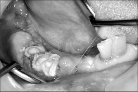
Fig. 2 Case 1. Necrotic bone was exposed on the mandible, right.
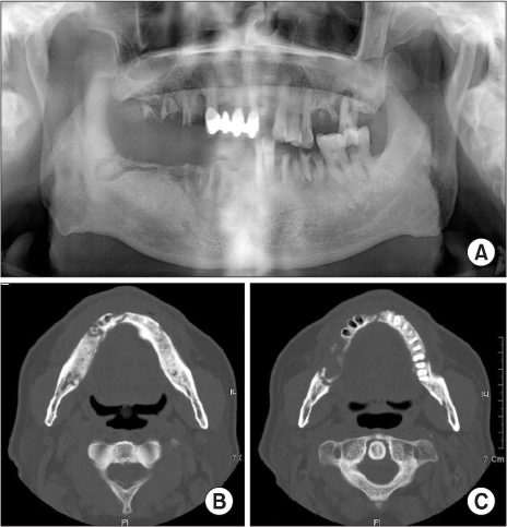
Fig. 3 Case 1. Preoperative radiographic findings. A. Large sequestrum was seen on the right mandible, and it was definitely distinguished from the surrounding bone on the orthopantograph. B. Mandibular computed tomography (CT) axial view; large sequestrum can be seen on the right mandible. C. Mandibular CT axial view; exfoliated socket can be seen on #33, 34, 35.
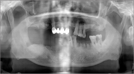
Fig. 4 Case 1. Post-operative radiographic findings post-operative day 8; the necrotic bone and multiple hopeless teeth were removed, and normal healing was noted.
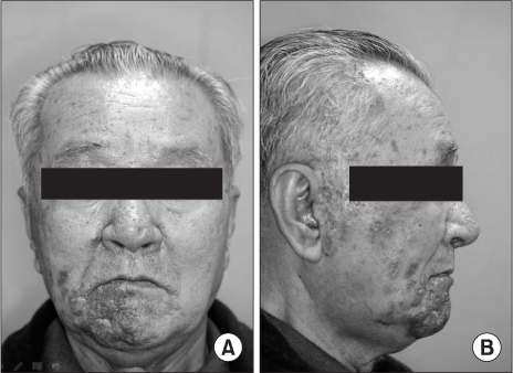
Fig. 5 Case 2. Skin lesions of herpes zoster; erythematous swelling and ulcer with post-inflammatory hyperpigmentation. A. Skin lesion can be seen on the chin, right. B. Skin lesion can be seen on the preauricular area, right.
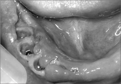
Fig. 6 Case 2. Necrotic bone was exposed, spontaneous teeth exfoliation on #43, 44, 45.
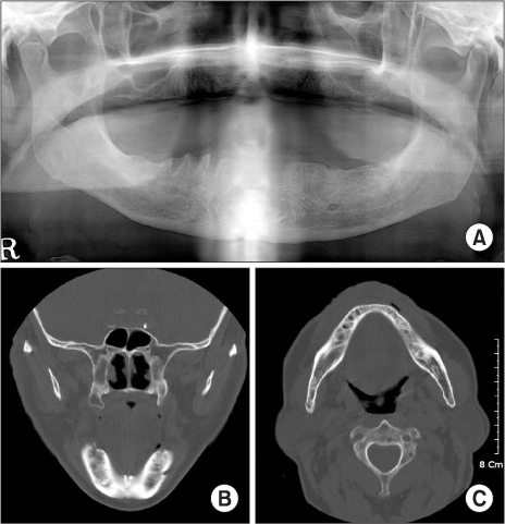
Fig. 7 Case 2. Radiographic findings; diffuse radiolucent lesion and extraction sockets area seen on the mandible, right. A. Orthopantograph. B. Mandibular computed tomography (CT) view: coronal. C. Mandibular CT view: axial.
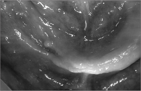
Fig. 8 Case 2. Soft tissue covering previous lesion.
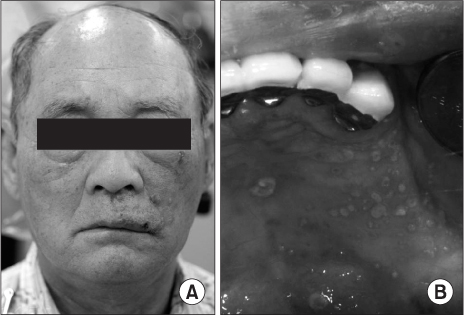
Fig. 9 Case 3. Lesions of herpes zoster (active phase). A. Extraoral view; diffuse erythematous plaques and vesicles were seen on the upper side of the face (V2 area) with tenderness. B. Intraoral view; multiple mucosal vesicles and ulcer formation observed on the maxilla, left.

Fig. 10 Case 3. Lesions of herpes zoster (healing phase). A. Extraoral view; clusters on the skin on the upper side of the face (V2 area) were seen. B. Intraoral view; normal healing state on mucosal lesion. No visible scar formation.
Reference
-
1. Gershon AA, Gershon MD, Breuer J, Levin MJ, Oaklander AL, Griffiths PD. Advances in the understanding of the pathogenesis and epidemiology of herpes zoster. J Clin Virol. 2010. 48:Suppl 1. S2–S7.
Article2. Mendieta C, Miranda J, Brunet LI, Gargallo J, Berini L. Alveolar bone necrosis and tooth exfoliation following herpes zoster infection: a review of the literature and case report. J Periodontol. 2005. 76:148–153.
Article3. Mintz SM, Anavi Y. Maxillary osteomyelitis and spontaneous tooth exfoliation after herpes zoster. Oral Surg Oral Med Oral Pathol. 1992. 73:664–666.
Article4. Jain MK, Manjunath KS, Jagadish SN. Unusual oral complications of herpes zoster infection: report of a case and review of literature. Oral Surg Oral Med Oral Pathol Oral Radiol Endod. 2010. 110:e37–e41.
Article5. Muto T, Tsuchiya H, Sato K, Kanazawa M. Tooth exfoliation and necrosis of the mandible--a rare complication following trigeminal herpes zoster: report of a case. J Oral Maxillofac Surg. 1990. 48:1000–1003.
Article6. Owotade FJ, Ugboko VI, Kolude B. Herpes zoster infection of the maxilla: case report. J Oral Maxillofac Surg. 1999. 57:1249–1251.
Article7. Rose F. Postherpetic neuralgia; trophic lesions in hand's bones similar to rheumatoid arthritis. Nouvelle Iconogr de la Salpêtrière. Soc Neurol. 1908. 9:64. [French].8. Dechaume M, Descrozailles C, Garlopeau F, Robert J. Localized mandibular necrosis during trigeminal herpes. Revue Stomatol. 1955. 56:516–521. [French].9. Schmader KE, Dworkin RH. Natural history and treatment of herpes zoster. J Pain. 2008. 9:1 Suppl 1. S3–S9.
Article10. Weaver BA. Herpes zoster overview: natural history and incidence. J Am Osteopath Assoc. 2009. 109:6 Suppl 2. S2–S6.11. Schwartz O, Kvorning SA. Tooth exfoliation, osteonecrosis of the jaw and neuralgia following herpes zoster of the trigeminal nerve. Int J Oral Surg. 1982. 11:364–371.
Article12. Sigurdsson A, Jacoway JR. Herpes zoster infection presenting as an acute pulpitis. Oral Surg Oral Med Oral Pathol Oral Radiol Endod. 1995. 80:92–95.
Article13. Ramchandani PL, Mellor TK. Herpes zoster associated with tooth resorption and periapical lesions. Br J Oral Maxillofac Surg. 2007. 45:71–73.
Article14. Arikawa J, Mizushima J, Higaki Y, Hoshino J, Kawashima M. Mandibular alveolar bone necrosis after trigeminal herpes zoster. Int J Dermatol. 2004. 43:136–137.
Article15. Garty BZ, Dinari G, Sarnat H, Cohen S, Nitzan M. Tooth exfoliation and osteonecrosis of the maxilla after trigeminal herpes zoster. J Pediatr. 1985. 106:71–73.
Article16. Wright WE, Davis ML, Geffen DB, Martin SE, Nelson MJ, Straus SE. Alveolar bone necrosis and tooth loss. a rare complication associated with herpes zoster infection of the fifth cranial nerve. Oral Surg Oral Med Oral Pathol. 1983. 56:39–46.
Article17. Hall HD, Jacobs JS, O'Malley JP. Necrosis of maxilla in patient with herpes zoster. Report of a case. Oral Surg Oral Med Oral Pathol. 1974. 37:657–662.18. Gilden D, Cohrs RJ, Mahalingam R, Nagel MA. aricella zoster virus vasculopathies: diverse clinical manifestations, laboratory features, pathogenesis, and treatment. Lancet Neurol. 2009. 8:731–740.
Article
- Full Text Links
-
- Actions
-
Cited
- CITED
-
- Close
- Share
-
- Similar articles
-
- A Clinical Case of Unilateral Maxillary Defect Reconstruction Using Nasolabial Flap
- Mandibular osteonecrosis following herpes zoster infection in the mandibular branch of the trigeminal nerve: a case report and literature review
- Acute Vestibular Neuritis Associated with Herpes Zoster Ophthalmicus
- Prominent Postinflammatory Hyperpigmentation with Herpes Zoster Ophthalmicus
- A Clinical Study of Herpes Zoster Ophthalmicus
