Congenital syngnathia: a case report
- Affiliations
-
- 1Department of Oral and Maxillofacial Surgery, College of Dentistry, Dankook University, Cheonan, Korea. kimchoms@dankook.ac.kr
- KMID: 2043847
- DOI: http://doi.org/10.5125/jkaoms.2012.38.3.171
Abstract
- Congenital syngnathia refers to the fusion of bony tissues, a rare disorder with only 41 cases reported in the international literature from 1936 to 2009. The occurrence of syngnathia without any other associated systemic disease or congenital anomaly is extremely rare. This report presents a case of congenital syngnathia with unilateral maxillomandibular bony adhesion without any other oral or maxillofacial anomaly. No recommended protocol for surgery exists due to the rarity of the disorder. There is a very low survival rate for the few patients who have forgone surgical management. This case describes a 74-year-old female patient who was suffering from limitation of mouth opening and was subsequently diagnosed with congenital syngnathia. The surgical staff performed separation surgery and reconstructed the malformed oral vestibule and cheek using the radial forearm free flap operation.
Keyword
MeSH Terms
Figure
-
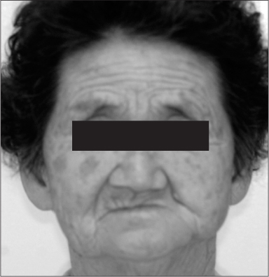
Fig. 1 74-year-old woman with facial asymmetry due to left mandibular hypoplasia resulting in the fusion of the left maxillo-mandibular alveolar ridge.

Fig. 2 Adhesion of the left commissural and retrocommissural oral cheek to the fused arches on the left side, which continued posteriorly as complete adhesion of the buccal mucosa to the slopes of the upper and lower arches, resulting in the absence of the buccal vestibule.

Fig. 3 Panoramic view showing left mandibular ramus hypoplasia resulting in the unilateral bony fusion of the left maxillary-mandibular alveolar ridges.

Fig. 4 A computerized 3 dimension reconstruction of the skull showed the bony fusion of the alveolar crest of the maxilla and mandible.
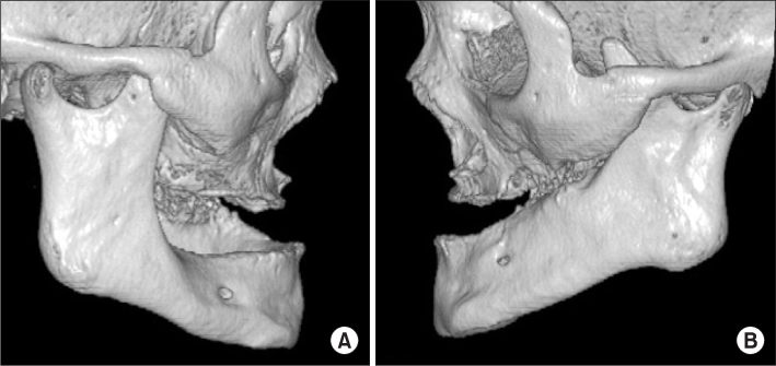
Fig. 5 Computerized 3 dimension reconstruction of the skull. A. There was neither temporomandibular joint (TMJ) ankylosis nor coronoid process hyperplasia in the right skull. B. There was no TMJ ankylosis, but coronoid process hyperplasia was noted on the left side.
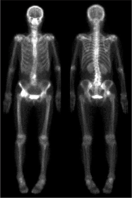
Fig. 6 The bone scan (99mTc-MDP) revealed a hot spot in the left maxillo-mandibular bony fusion area.
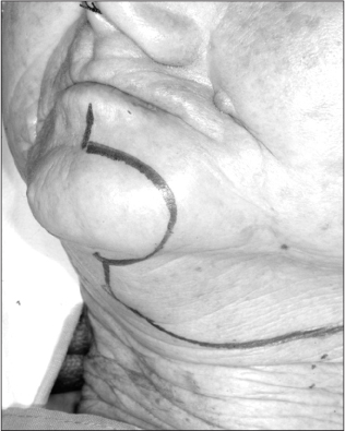
Fig. 7 Surgical incision. The incision line was lip-split and extended to apron flap incision.
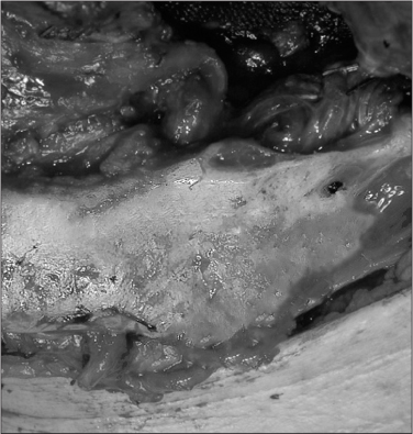
Fig. 8 The maxillo-mandibular fusion area is separated by a saw, and left coronoidectomy was done to favor mouth opening training.
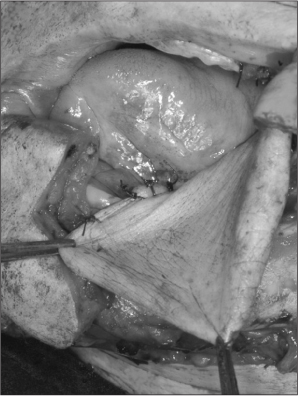
Fig. 9 Radial forearm free flap was inserted for the reconstruction of the buccal cheek mucosa.
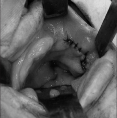
Fig. 10 Debulking procedure for the reduction of volume of excessive radial forearm free flap during the second operation. Mouth opening of 45 mm was achieved.
Reference
-
1. Naikmasur VG, Sattur AP, Joshi SK, Rai A. Congenital syngnathia: case report and review of literature. Cleft Palate Craniofac J. 2010. 47:654–660.
Article2. Burket LW. Congenital bony temporomandibular ankylosis and facial hemiatrophy. JAMA. 1936. 106:1719–1722.
Article3. Dawson KH, Gruss JS, Myall RW. Congenital bony syngnathia: a proposed classification. Cleft Palate Craniofac J. 1997. 34:141–146.
Article4. Laster Z, Temkin D, Zarfin Y, Kushnir A. Complete bony fusion of the mandible to the zygomatic complex and maxillary tuberosity: case report and review. Int J Oral Maxillofac Surg. 2001. 30:75–79.
Article5. Poswillo D. The pathogenesis of the first and second branchial arch syndrome. Oral Surg Oral Med Oral Pathol. 1973. 35:302–328.
Article6. Hegtvedt AK. Peterson LJ, editor. Diagnosis and management of facial asymmetry. Principles of oral and maxillofacial surgery. 1992. Philadelphia: Lippincott;1400–1401.7. Rao S, Oak S, Wagh M, Kulkarni B. Congenital midline palato-mandibular bony fusion with a mandibular cleft and a bifid tongue. Br J Plast Surg. 1997. 50:139–141.
Article8. Kamata S, Satoh K, Uemura T, Onizuka T. Congenital bilateral zygomatico-mandibular fusion with mandibular hypoplasia. Br J Plast Surg. 1996. 49:251–253.
Article9. Goodacre TE, Wallace AF. Congenital alveolar fusion. Br J Plast Surg. 1990. 43:203–209.
Article10. Nanda R. Maxillomandibular ankylosis and cleft palate in rat embryos. J Dent Res. 1970. 49:1086–1090.
Article11. Humphrey T. The relation between human fetal mouth opening reflexes and closure of the palate. Am J Anat. 1969. 125:317–344.
Article12. Subramanian B, Agrawal K, Panda K. Congenital fusion of the jaws: a management protocol. Int J Oral Maxillofac Surg. 2010. 39:925–929.
Article13. Agrawal K, Chandra SS, Sreekumar NS. Congenital bilateral intermaxillary bony fusion. Ann Plast Surg. 1993. 30:163–166.
Article14. Choi JY, Min CG, Myoung H, Hwang SJ, Kim MJ, Lee JH. Acquired syngnathia. Br J Oral Maxillofac Surg. 2004. 42:448–450.
Article
- Full Text Links
-
- Actions
-
Cited
- CITED
-
- Close
- Share
-
- Similar articles
-
- Isolated Congenital Alveolar Synechiae: Review of Literature and Case Report: A Case Report
- AIRWAY MANAGEMENT FOR SYNGNATHIA
- Familial Congenital Muscular Torticollis: A Case Report
- Congenital self-healing reticulohistiocytosis: report of a case of the solitary type and review of the literature
- A Case of Congenital Hydronephrosis
